. 2012 May 4;287(19):15544-56.
doi: 10.1074/jbc.M111.330365. Epub 2012 Mar 12.

Mononuclear iron enzymes are primary targets of hydrogen peroxide stress

Affiliations

Mononuclear iron enzymes are primary targets of hydrogen peroxide stress

Adil Anjem et al. J Biol Chem. .

Abstract

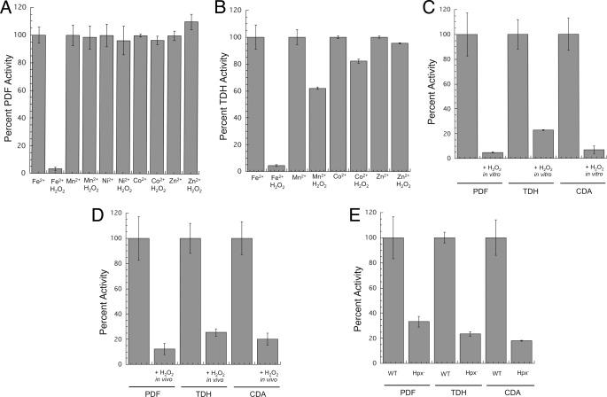
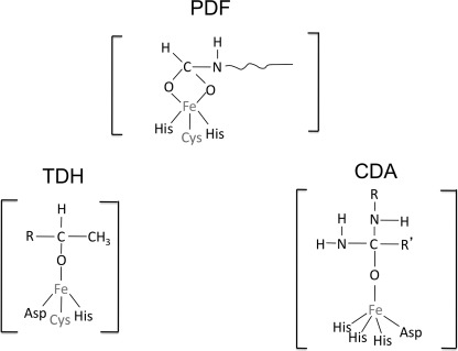
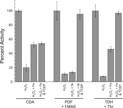
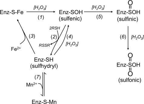
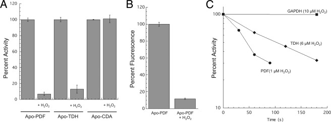
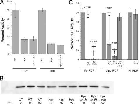
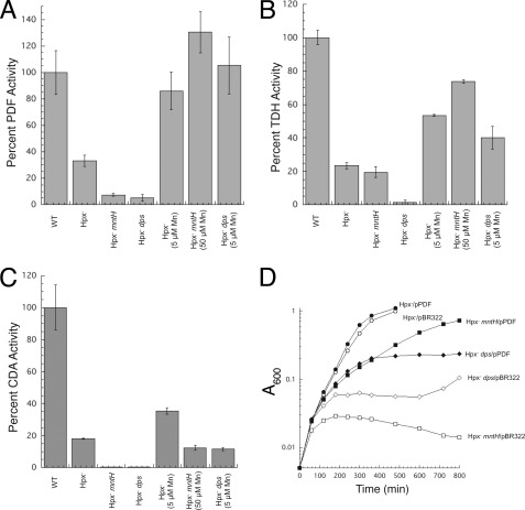
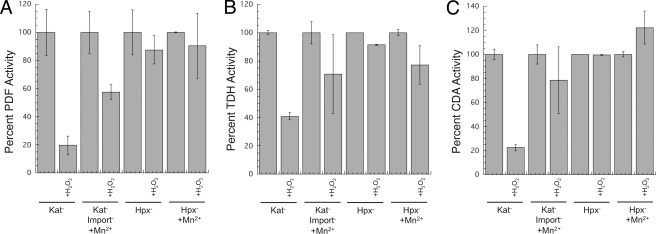
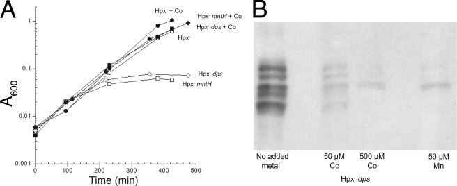
This study tested whether nonredox metalloenzymes are commonly charged with iron in vivo and are primary targets of oxidative stress because of it. Indeed, three sample mononuclear enzymes, peptide deformylase, threonine dehydrogenase, and cytosine deaminase, were rapidly damaged by micromolar hydrogen peroxide in vitro and in live Escherichia coli. The first two enzymes use a cysteine residue to coordinate the catalytic metal atom; it was quantitatively oxidized by the radical generated by the Fenton reaction. Because oxidized cysteine can be repaired by cellular reductants, the effect was to avoid irreversible damage to other active-site residues. Nevertheless, protracted H(2)O(2) exposure gradually inactivated these enzymes, consistent with the overoxidation of the cysteine residue to sulfinic or sulfonic forms. During H(2)O(2) stress, E. coli defended all three proteins by inducing MntH, a manganese importer, and Dps, an iron-sequestration protein. These proteins appeared to collaborate in replacing the iron atom with nonoxidizable manganese. The implication is that mononuclear metalloproteins are common targets of H(2)O(2) and that both structural and metabolic arrangements exist to protect them.


Figures

FIGURE 1.
FIGURE 1.
PDF, TDH, and CDA are sensitive to H2O2 stress in vitro and in vivo. A, purified PDF was metallated with various transition metals (100 μm) and then challenged with H2O2 (10 μm for 5 min). B, purified TDH was metallated with various transition metals (100 μm) and then challenged with H2O2 (10 μm for 5 min). C, as-isolated PDF, TDH, and CDA activities of crude extracts were sensitive to H2O2. Catalase-deficient (ΔkatE ΔkatG) strains were grown in aerobic medium, and extracts were exposed to 10 μm H2O2 for 5 min. D, enzymes are sensitive to a bolus of H2O2 in vivo. Catalase-deficient (ΔkatE ΔkatG) strains were grown in aerobic medium and briefly exposed to H2O2 (100 μm for 10 min). Exogenous metal was added to the assay to ensure full activity of undamaged enzymes, as described under “Experimental Procedures.” E, enzyme activities are low in cells that lack scavenging enzymes. Wild-type (MG1655) and Hpx (LC106) strains were grown in aerobic medium, and enzyme activities were measured.
FIGURE 2.
FIGURE 2.
Metal-substrate interactions within the actives sites of PDF (35), TDH (60), and CDA (61).
FIGURE 3.
FIGURE 3.
H2O2 stress reversibly oxidizes the cysteine residues of iron-charged PDF and TDH. Iron-charged enzymes were exposed to 20 μm H2O2 for 5 min. After the addition of catalase, enzyme activity was measured before and after the addition of iron (CDA and TDH) or nickel (PDF) and of TCEP.
FIGURE 4.
FIGURE 4.
Proposed pathways for the damage and reactivation of PDF and TDH.
FIGURE 5.
FIGURE 5.
Apo-PDF and apo-TDH can be directly oxidized by H2O2. A, purified apo-PDF and apo-TDH and apo-CDA in cell extracts were exposed to 0 or 10 μm H2O2 for 5 min. After addition of catalase, enzymes were reconstituted with iron (CDA), zinc (TDH), or nickel (PDF) and assayed. B, direct measurement of sulfhydryl oxidation in PDF. Purified apo-PDF was challenged with zero or 20 μm H2O2 for 1 min. The enzyme was then treated with monobromobimane, and fluorescence was measured. C, comparison of the sensitivities of apo-PDF, apo-TDH, and GAPDH to H2O2. Pure apo-PDF and pure apo-TDH were challenged with 1 and 6 μm H2O2, respectively, at RT prior to remetallation and assay. The sensitivity of GAPDH to 10 μm H2O2 was measured in cell extracts (JI367, ΔkatE ΔkatG). Extracts did not protect PDF or TDH (data not shown).
FIGURE 6.
FIGURE 6.
Extended exposure to H2O2 converts PDF and TDH to nonreactivatible forms in vivo and in vitro. A, TCEP did not restore PDF and TDH activities in the extracts of Hpx cells that had been grown in aerobic medium. Both enzymes were assayed in the presence of metal (500 μm Ni2+ and 500 μm Fe2+ respectively) to ensure full activity of undamaged enzymes. B, PDF polypeptide was not degraded in vivo. Anaerobic cells expressing PDF-FLAG were treated with chloramphenicol and aerated starting at time 0. Polypeptide content was monitored by Western blot. The strains used were MG1655/pPDF-FLAG, LC106/pPDF-FLAG (Hpx) and AA30/pPDF-FLAG (Hpx ΔmntH). C, iron-PDF and apo-PDF can be overoxidized in vitro. Iron-charged PDF, apo-PDF, and nickel-charged PDF were exposed to H2O2 (133, 100, and 500 μm, respectively) for the indicated times. They were then treated ± TCEP and reconstituted with nickel prior to assay.
FIGURE 7.
FIGURE 7.
Manganese import (by MntH) and iron sequestration (by Dps) are key elements in protecting PDF, TDH, and CDA from H2O2. A–C, PDF, TDH, and CDA activities in wild-type (MG1655, WT), Hpx (LC106), Hpx ΔmntH (AA30), and Hpx Δdps (SP66) strains. Where indicated, extra manganese was included in the growth medium. Assays were conducted after anaerobic reconstitution with nickel (PDF) or iron (TDH and CDA). Time courses of PDF and TDH inactivation are shown as supplemental Fig. S2. D, overexpression of PDF relieves the Hpx ΔmntH growth defect. At time 0, anaerobic cultures were diluted into aerobic medium. The strains used were LC106 (Hpx, circles), AA30 (Hpx ΔmntH, squares), and SP66 (Hpx Δdps, diamonds). Closed symbols, plasmid overexpressing PDF; open symbols, vector control.
FIGURE 8.
FIGURE 8.
Intracellular manganese protects PDF (A), TDH (B), and CDA (C) from a bolus of H2O2. Cells were grown in aerobic medium that was supplemented with 5 μm MnCl2 where indicated. Chloramphenicol was added, and cells were exposed to 100 μm H2O2 for 10 min. Enzyme activities were measured in extracts after metallation with nickel (PDF) or iron (TDH and CDA). Strains used are as follows: JI367 (Kat), AA301 (Kat import), and LC106 (Hpx).
FIGURE 9.
FIGURE 9.
Cobalt can substitute for manganese in protecting H2O2-stressed cells. A, cobalt supplements restore the growth of both Hpx ΔmntH and Hpx Δdps cells. At time 0, anaerobic cultures were diluted into aerobic medium. Where indicated, 30 μm CoCl2 was included (closed symbols). Strains used are as follows: LC106 (Hpx, circles), AA30 (Hpx ΔmntH, squares), and SP66 (Hpx Δdps, diamonds). B, cobalt suppresses protein carbonylation in the Hpx Δdps cells.

References

    1. Gort A. S., Imlay J. A. (1998) Balance between endogenous superoxide stress and antioxidant defenses. J. Bacteriol. 180, 1402–1410 - PMC - PubMed
    1. Seaver L. C., Imlay J. A. (2001) Hydrogen peroxide fluxes and compartmentalization inside growing Escherichia coli. J. Bacteriol. 183, 7182–7189 - PMC - PubMed
    1. Delaunay A., Pflieger D., Barrault M. B., Vinh J., Toledano M. B. (2002) A thiol peroxidase is an H2O2 receptor and redox-transducer in gene activation. Cell 111, 471–481 - PubMed
    1. Choi H., Kim S., Mukhopadhyay P., Cho S., Woo J., Storz G., Ryu S. (2001) Structural basis of the redox switch in the OxyR transcription factor. Cell 105, 103–113 - PubMed
    1. Lee J. W., Helmann J. D. (2006) The PerR transcription factor senses H2O2 by metal-catalyzed histidine oxidation. Nature 440, 363–367 - PubMed

Publication types

MeSH terms

LinkOut - more resources