. 2012;10(3):e1001281.
doi: 10.1371/journal.pbio.1001281. Epub 2012 Mar 6.

Two programmed cell death systems in Escherichia coli: an apoptotic-like death is inhibited by the mazEF-mediated death pathway

Affiliations

Two programmed cell death systems in Escherichia coli: an apoptotic-like death is inhibited by the mazEF-mediated death pathway

Ariel Erental et al. PLoS Biol. 2012.

Abstract

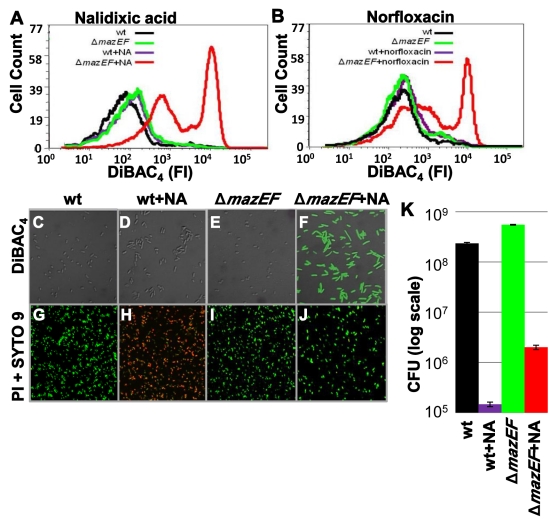
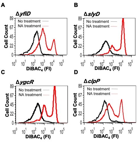
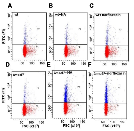
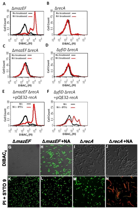
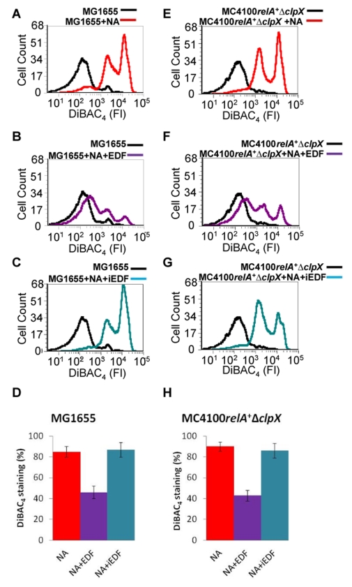
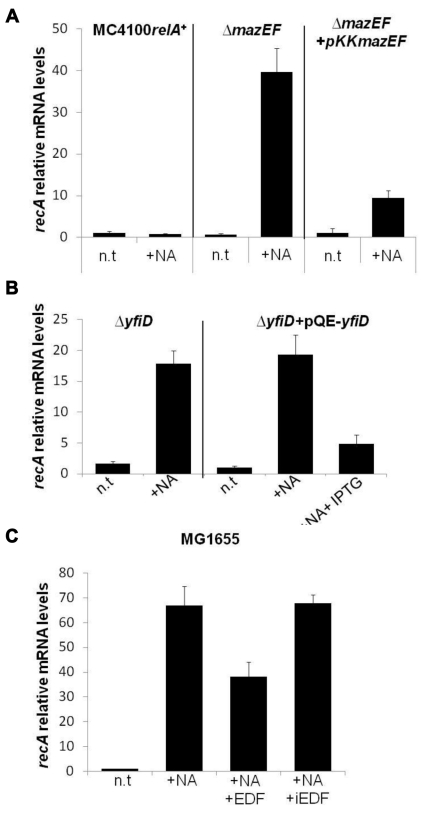
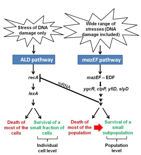
In eukaryotes, the classical form of programmed cell death (PCD) is apoptosis, which has as its specific characteristics DNA fragmentation and membrane depolarization. In Escherichia coli a different PCD system has been reported. It is mediated by the toxin-antitoxin system module mazEF. The E. coli mazEF module is one of the most thoroughly studied toxin-antitoxin systems. mazF encodes a stable toxin, MazF, and mazE encodes a labile antitoxin, MazE, which prevents the lethal effect of MazF. mazEF-mediated cell death is a population phenomenon requiring the quorum-sensing pentapeptide NNWNN designated Extracellular Death Factor (EDF). mazEF is triggered by several stressful conditions, including severe damage to the DNA. Here, using confocal microscopy and FACS analysis, we show that under conditions of severe DNA damage, the triggered mazEF-mediated cell death pathway leads to the inhibition of a second cell death pathway. The latter is an apoptotic-like death (ALD); ALD is mediated by recA and lexA. The mazEF-mediated pathway reduces recA mRNA levels. Based on these results, we offer a molecular model for the maintenance of an altruistic characteristic in cell populations. In our model, the ALD pathway is inhibited by the altruistic EDF-mazEF-mediated death pathway.


Conflict of interest statement

The authors have declared that no competing interests exist.

Figures

Figure 1
Figure 1. The effect of DNA damage on membrane depolarization and viability.
The effect on membrane depolarization (detected by DiBAC4) (A–F), cell viability (detected by Live/Dead Kit) (G–J), and CFU (K). E. coli strains MC4100relA + (WT) or MC4100relA +ΔmazEF were grown to OD600 0.6. Each culture was divided into two aliquots, and then the appropriate antibiotics were added to one of the samples: (A) NA (100 µg/ml) or (B) norfloxacin (1.5 µg/ml). Cells were incubated for 4 h and then stained with DiBAC4. (A and B) The intensity of the fluorescence (FI) of DiBAC4 in the cells was determined by FACS. The fraction of cells in which membrane depolarization occurred was determined as being above 103 DiBAC4. (C–F) The fluorescence intensity of DiBAC4 in the cells was also determined by confocal microscopy. (G–J) Confocal microscopy of the cells treated with NA as in (C–F), but stained with the Live-Dead Kit. (K) The effect of NA on CFU. All data are representative of three independent experiments.
Figure 2
Figure 2. Deleting genes acting downstream from mazF leads to membrane depolarization.
Cultures of WT E. coli MC4100relA + and of derivative strains from which we deleted (A) yfiD, (B) slyD, (C) ygcR, and (D) clpP were grown, treated with NA (100 µg/ml), and stained with DiBAC4 as described in Figure 1. Samples treated with NA are shown in red, and samples without treatment are shown in black. FI, fluorescence intensity.
Figure 3
Figure 3. The effect of DNA damage on DNA fragmentation (detected by TUNEL assay).
E. coli (A–C) MC4100relA + (WT) or (D–F) MC4100relA +ΔmazEF were grown to OD600 0.6. Each culture was divided into three samples: (A and D) without treatment; (B and E) 100 µg/ml NA was added, and (C and F) 1.5 µg/ml norfloxacin was added. DNA fragmentation was determined using the Apo-Direct Kit. The location of the horizontal threshold line between TUNEL negative cells (highlighted in red) and TUNEL positive cells (highlighted in blue) was determined based on untreated and unstained MC4100relA + cells. All data are representative of three independent experiments. FI, fluorescence intensity; FSC, forward scatter.
Figure 4
Figure 4. The effect of DNA damage on membrane depolarization is recA-dependent.
(A–D) E. coli (A) MC4100relA +ΔmazEF, (B) MC4100relA +ΔrecA, (C) MC4100relA +ΔmazEFΔrecA, and (D) MC4100relA +ΔyfiDΔrecA cells were grown, treated with NA (100 µg/ml), and stained with DiBAC4 as in Figure 1. Samples treated with NA are shown in red. Control samples without treatment are shown in black. (E and F) Complementation assay with plasmid pQE32-recA. E. coli strains (E) MC4100relA +ΔmazEFΔrecA and (F) MC4100relA +ΔyfiDΔrecA harboring pQE32-recA were grown and treated with NA (100 µg/ml) and in the presence (red) or absence (black) of 1 mM IPTG. The cultures were stained with DiBAC4, and the fluorescence intensity (FI) was determined by FACS as described in Figure 1. (G–J) The fluorescence intensity of DiBAC4 in cells of MC4100relA +ΔmazEF and MC4100relA +ΔrecA that were treated with NA was also determined by confocal microscopy. (K–N) Confocal microscopy of the cells treated with NA as in (G–J), but stained with the Live/Dead Kit. All data are representative of three independent experiments.
Figure 5
Figure 5. The effect of EDF on membrane depolarization.
After the various E. coli strains were grown to OD600 0.6, the cells were treated as follows: (A) MG1655+100 µg/ml NA; (B) MG1655+100 µg/ml NA+50 ng/ml EDF; (C) MG1655+100 µg/ml NA+50 ng/ml iEDF (NNGNN); (E) MC4100relA +ΔclpX+100 µg/ml NA; (F) MC4100relA +ΔclpX+100 µg/ml NA+200 ng/ml EDF; (G) MC4100relA +ΔclpX+100 µg/ml NA+200 ng/ml iEDF. The quantitative effect of NA and NA+EDF on the percent of DiBAC4-stained cells is shown in (D) (data from [A–C]) and (H) (data from [E–G]). Error bars in (D) and (H) indicate standard deviation. Staining, FACS analysis, and determining the fraction of the DiBAC4-stained (membrane depolarized) cells were carried out as described in the legend to Figure 1. All data are representative of three independent experiments. FI, fluorescence intensity.
Figure 6
Figure 6. The mazEF pathway reduces the levels of recA mRNA.
(A) E. coli cells of strain MC4100relA +, its ΔmazEF derivative, or ΔmazEF carrying plasmid pKKmazEF were grown and treated with NA (100 µg/ml) or left without treatment (n.t) as in Figure 1. Then RNA was extracted from the cells, and real-time PCR was performed to quantify recA mRNA levels. The indicated values are relative to recA RNA levels in untreated MC4100relA + cells. (B) E. coli MC4100relA +ΔyfiD or E. coli MC4100relA +ΔyfiD cells carrying the inducible plasmid pQE-yfiD were grown and treated with NA as in (A). To induce the expression of yfiD, 1 mM of IPTG was added before the addition of NA. The indicated values are relative to recA RNA levels in untreated MC4100relA +ΔyfiD cells. (C) E. coli MG1655 cells were grown and treated with NA (100 µg/ml) or left without treatment as in (A). To test the effect of EDF on this strain, a synthetic EDF (50 ng/ml) or iEDF (50 ng/ml) (NNGNN) was added to the cells before adding the NA. The indicated values are relative to recA RNA levels in untreated MG1655 cells. All experiments were performed in triplicate, and a typical experiment out of three is shown. Error bars indicate standard deviation.
Figure 7
Figure 7. A suggested model for the inhibition of the recA- and lexA-mediated apoptotic-like pathway by the EDF-mazEF pathway.
Our model is based on the mazEF downstream pathway and the EDF-mazEF-mediated cell death pathway ,– that we have described previously ,– and on the recA- and lexA-mediated apoptotic-like pathway on which we have reported here. For additional explanations see also Results and Discussion.

Comment in

References

    1. Jacobson M. D, Weil M, Raff M. C. Programmed cell death in animal development. Cell. 1997;88:347–354. - PubMed
    1. Nagata A. Apoptosis by death factor. Cell. 1997;88:355–365. - PubMed
    1. Kerr J. F. R, Wyllie A. H, Curie A. R. Apoptosis: a basic biological phenomenon with wide-ranging implications in tissue kinetics. Br J Cancer. 1972;4:239–257. - PMC - PubMed
    1. Hengartner M. O. The biochemistry of apoptosis. Nature. 2009;407:770–776. - PubMed
    1. Hotchkiss R. S, Strasser A, McDunn J. E, Swanson P. E. Cell death. N Engl J Med. 2009;361:1570–1583. - PMC - PubMed

Publication types

MeSH terms