. 2012;7(3):e31823.
doi: 10.1371/journal.pone.0031823. Epub 2012 Mar 7.

Prolyl isomerase Pin1 regulates mouse embryonic fibroblast differentiation into adipose cells

Affiliations

Prolyl isomerase Pin1 regulates mouse embryonic fibroblast differentiation into adipose cells

Takafumi Uchida et al. PLoS One. 2012.

Abstract

Background: A peptidyl prolyl cis/trans isomerase, Pin1, regulates insulin signal transduction. Pin1 reduces responses to insulin stimulation by binding CRTC2 (CREB-regulated transcriptional co-activator 2) and PPARγ (peroxisome prolifereator- activated receptor γ), but conversely enhances insulin signaling by binding IRS-1 (insulin receptor substrate-1), Akt kinase, and Smad3. Therefore, it is still unclear whether Pin1 inhibits or enhances adipose cell differentiation.

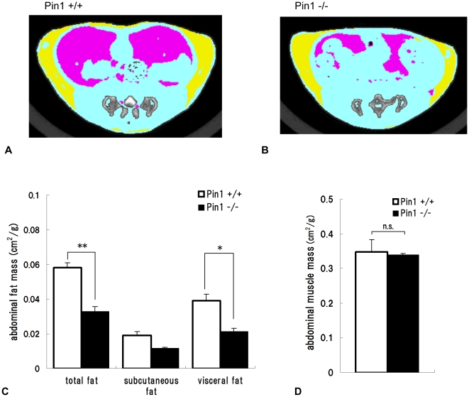
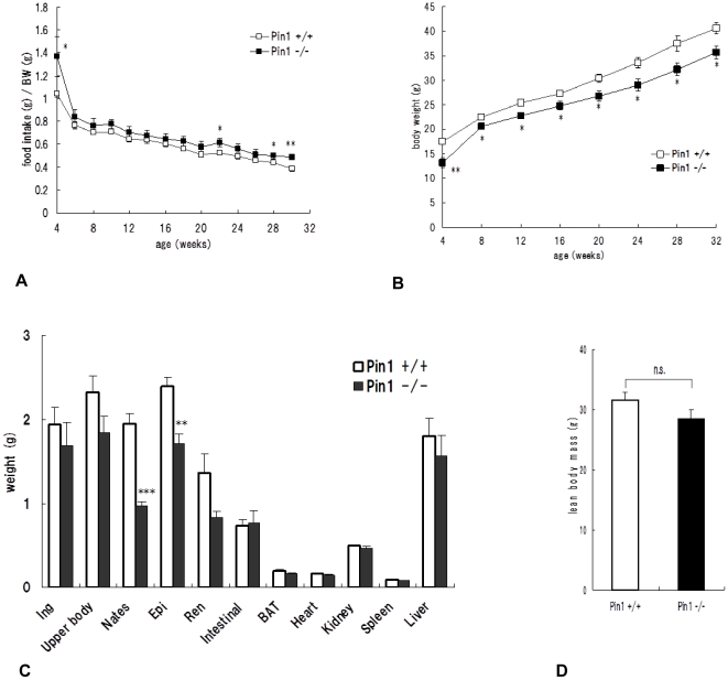
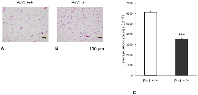
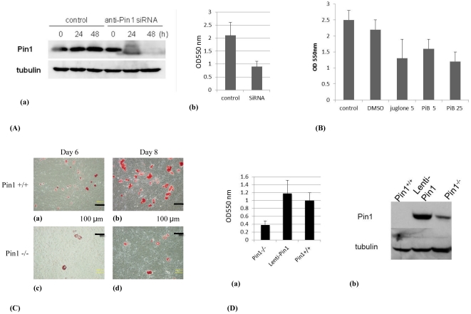
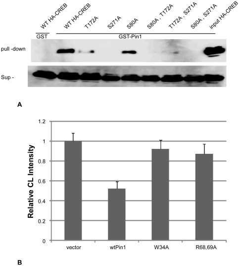
Methodology/principal findings: Pin1(-/-) and wild-type mice were fed with high fat diets and adipose tissue weight was measured. Compared to wild-type mice, Pin1(-/-) mice had lower adipose tissue weight, while the weight of other tissues was similar. Mouse embryo fibroblasts (MEFs), prepared from both groups of mice, were induced to differentiate into adipose cells by stimulation with insulin. However, the rate of differentiation of MEFs from Pin1(-/-) mice was less than that of MEFs from wild-type mice. The rate of insulin-induced MEF cell differentiation in Pin1(-/-) mice was restored by increasing expression of Pin1. We found that Pin1 binds to phosphoThr172- and phosphoSer271-Pro sites in CREB suppress the activity in COS-7 cells.

Conclusion and significance: Pin1 enhanced the uptake of triglycerides and the differentiation of MEF cells into adipose cells in response to insulin stimulation. Results of this study suggest that Pin1 down-regulation could be a potential approach in obesity-related dysfunctions, such as high blood pressure, diabetes, non-alcoholic steatohepatitis.


Conflict of interest statement

Competing Interests: The authors have declared that no competing interests exist.

Figures

Figure 1
Figure 1. Computed Tomography analysis of wild-type and Pin1−/− mice.
The abdomens of 16 week-old male wild-type (A) and Pin1−/− (B) mice were scanned (yellow; subcutaneous fat, pink; visceral fat, blue; muscle). Quantities of subcutaneous, visceral, and total adipose tissues (**p<0.01) (C), and the other tissues, mainly muscle (D), were measured from the images. The areas were measured from the computer tomography images (cm2) and they were adjusted for body weights of mice (±SEM, n = 3).
Figure 2
Figure 2. Relationship between quantity of fat and Pin1 expression.
High fat diets were fed to wild-type (n = 8) and Pin1−/− (n = 5) mice between the ages of 4–28 weeks and the amount of food intake (A) and body weight (B) were recorded. Weights of subcutaneous and genital fat tissues were measured (C). The total weight of all fat tissues removed was also measured (D). The amount of food intake per mouse weight was monitored. Student t-test *p<0.05, **p<0.01, ***p<0.001.
Figure 3
Figure 3. Pathological analysis of adipose tissues from wild-type and Pin1−/− mice.
Paraffin-embedded sections of inguinal fat tissue from wild-type (A) and Pin1−/− (B) mice were stained with hematoxylin and eosin. The size of cells was analyzed quantitatively with Image J analysis soft. The average sizes of the adipose cells of wild and Pin1−/− mice were 6129.6±136.0 µm2 and 3516.6±87.0 µm2 (±SEM) respectively (Figure 3C). Student t-test p<0.001.
Figure 4
Figure 4. Differentiation of fibroblasts into adipose cells.
(A) NIH3T3L1 cells were transfected with 20 µg mock or SiRNA-Pin1 plasmids, cultured for 24 and 48 hours, and the expression levels of Pin1 were examined with western blot analysis (a). The NIH3T3-L1 cells were incubated in DMEM medium, containing; 0.5 mM 3-isobutyl-1-methylxanthine, 1 µM dexamethasone, and 1.7 µM insulin for 48 hours, treated with 4% paraformaldehyde and 60% 2-propanol and then stained with Oil Red O. The amount of Oil Red O extracted from cells was determined by measuring the absorbance at 550 nm (b). (B) NIH3T3-L1 cells were incubated in the same medium with juglone (5 µM) or PiB (5, 25 µM), and the fat was measured with oil red O assay. (C) Wild-type (a, b) and Pin1−/− (c, d) MEFs were stimulated in the same way as NIH3T3 cells for 6 (a, c) and 8 days (b, d), stained with Oil Red O, and analyzed by microscopy. (D) Pin1−/− MEF, Lenti-Pin1 (Pin1−/− MEF-infected with lentiviral Pin1 cDNA), and wild-type MEF were also examined like (C) (a). Pin1 levels of the Pin1−/− MEFs, Lenti-Pin1−/−, and wild-type MEFs were analyzed with western blot (b).
Figure 5
Figure 5. Association of Pin1 and CREB.
(A) Lysates of COS-7 cells, transfected with HA-CREB (wild-type; T172A; S271A; S80A; S80A/T172A; T172A/S271A; S80A/S271A) and stimulated with 10 µM forskolin for 6 hr, were pulled down with GST-Sepharose (control), GST-Pin1-Sepharose, and input (upper panel). CREB levels in the supernatant are shown in the lower panel. (B) 106 of COS-7 were co-transfected with pCRE-Luc and CMV-Pin1 (wt; wild type, W34A; mutation at WW domain, R68,69A; prolyl isomerase mutant). The cells were treated with 100 µM of forskolin (CST) for 6 hours and luciferase activities were determined using the Dual-Luciferase Reporter Assay System (Promega).

References

    1. Fujimori F, Takahashi K, Uchida C, Uchida T. Mice lacking Pin1 develop normally, but are defective in entering cell cycle from G0 arrest. Biochem Biophys Res Commun. 1999;265:658–663. - PubMed
    1. Liou YC, Ryo A, Huang HK, Lu PJ, Bronson R, et al. Loss of Pin1 function in the mouse resembles the cyclin D1- null phenotypes. Proc Natl Aca Sci USA. 2002;99:1335–1340. - PMC - PubMed
    1. Fujimoto Y, Shiraki T, Horiuchi Y, Waku T, Shigenaga A, et al. Proline cis/trans-Isomerase Pin1 Regulates Peroxisome Proliferator-activated Receptor γ Activity through the Direct Binding to the Activation Function-1 Domain. J Biol Chem. 2010;285:3126–3132. - PMC - PubMed
    1. Nakatsu Y, Sakoda H, Kushiyama A, Ono H, Fujishiro M, et al. Pin1 associates with and induces translocation of CRTC2 to the cytosol, thereby suppressing CRE transcriptional activity. J Biol Chem. 2010;285:33018–33027. - PMC - PubMed
    1. Nakatsu Y, Sakoda H, Kushiyama A, Zhang J, Ono H, et al. Peptidyl-prolyl cis/trans isomerase NIMA-interacting 1 associates with IRS-1 and enhances insulin actions and adipogenesis. J Biol Chem. 2011;286:20812–20822. - PMC - PubMed

Publication types

MeSH terms

Substances