. 2012;7(3):e32526.
doi: 10.1371/journal.pone.0032526. Epub 2012 Mar 8.

Aronia melanocarpa juice induces a redox-sensitive p73-related caspase 3-dependent apoptosis in human leukemia cells

Affiliations

Aronia melanocarpa juice induces a redox-sensitive p73-related caspase 3-dependent apoptosis in human leukemia cells

Tanveer Sharif et al. PLoS One. 2012.

Abstract

Polyphenols are natural compounds widely present in fruits and vegetables, which have antimutagenic and anticancer properties. The aim of the present study was to determine the anticancer effect of a polyphenol-rich Aronia melanocarpa juice (AMJ) containing 7.15 g/L of polyphenols in the acute lymphoblastic leukemia Jurkat cell line, and, if so, to clarify the underlying mechanism and to identify the active polyphenols involved. AMJ inhibited cell proliferation, which was associated with cell cycle arrest in G(2)/M phase, and caused the induction of apoptosis. These effects were associated with an upregulation of the expression of tumor suppressor p73 and active caspase 3, and a downregulation of the expression of cyclin B1 and the epigenetic integrator UHRF1. AMJ significantly increased the formation of reactive oxygen species (ROS), decreased the mitochondrial membrane potential and caused the release of cytochrome c into the cytoplasm. Treatment with intracellular ROS scavengers prevented the AMJ-induced apoptosis and upregulation of the expression of p73 and active caspase 3. The fractionation of the AMJ and the use of identified isolated compounds indicated that the anticancer activity was associated predominantly with chlorogenic acids, some cyanidin glycosides, and derivatives of quercetin. AMJ treatment also induced apoptosis of different human lymphoblastic leukemia cells (HSB-2, Molt-4 and CCRF-CEM). In addition, AMJ exerted a strong pro-apoptotic effect in human primary lymphoblastic leukemia cells but not in human normal primary T-lymphocytes. Thus, the present findings indicate that AMJ exhibits strong anticancer activity through a redox-sensitive mechanism in the p53-deficient Jurkat cells and that this effect involves several types of polyphenols. They further suggest that AMJ has chemotherapeutic properties against acute lymphoblastic leukemia by selectively targeting lymphoblast-derived tumor cells.


Conflict of interest statement

Competing Interests: The authors have declared that no competing interests exist.

Figures

Figure 1
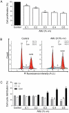
Figure 1. AMJ caused a concentration-dependent inhibition of cell proliferation and induction of G2/M cell cycle arrest in Jurkat cells.
Cells were exposed to increasing concentrations of AMJ for 24 h. (A) cell proliferation rate was determined using MTS assay. The absolute value obtained for each AMJ-treated sample is expressed as percent relative to the absolute value obtained for the untreated sample and set at 100%. (B) and (C) cell cycle distribution was assessed by flow cytometry using the DNA fluorochrome PI detection assay. (B) shows representative DNA content histograms for treated (right panel) or untreated (left panel) cells. (C) Corresponding cumulative data. The number of cells in each mitosis phase is determined and expressed as percent relative to the total cell number. Values are shown as means ± S.E.M. (n = 3); *, P<0.05 versus respective control.
Figure 2
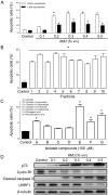
Figure 2. AMJ and AMJ-derived fractions induced a pro-apoptotic response in Jurkat cells.
Cells were exposed either to increasing concentrations of AMJ, or AMJ-derived fractions (tested at 21.4 µg/mL, equivalent to the polyphenol content of AMJ 0.3% v/v), or various isolated pure products (100 µM) for 24 h. (A), (B) and (C) cell apoptosis rates were assessed by flow cytometry using the annexin V-FITC/PI apoptosis assay. (D) The expression level of key regulators of cell cycle and apoptosis was assessed by Western blot analysis. The data are representative of at least three independent experiments. Values are shown as means ± S.E.M. (n = 3); *, P<0.05 versus respective control.
Figure 3
Figure 3. AMJ induced a pro-oxidant response and altered the ΔΨm in Jurkat cells.
Cells were exposed to increasing concentrations of AMJ for 24 h. (A) and (B) The formation of ROS was assessed by flow cytometry after incubation with the redox-sensitive fluorescent probe DHE. Cells were exposed to antioxidant treatment as indicated 30 min before the addition of AMJ. (C) Changes in the ΔΨm were assessed by flow cytometry using the fluorescent probe DiOC6. Values are shown as means ± S.E.M. (n = 3); *, P<0.05 versus control; †, P<0.05 versus AMJ treatment. (D) The level of cytochrome c in the cytosolic fractions of Jurkat cells was assessed by Western blot analysis. Similar findings were observed in three independent experiments.
Figure 4
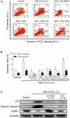
Figure 4. AMJ induced a ROS-dependent pro-apoptotic effect in Jurkat cells.
Cells were exposed to antioxidant treatment as indicated 30 min before the addition of AMJ for 24 h. (A) and (B) cell apoptosis rates were assessed by flow cytometry using the annexin V-FITC/PI apoptosis assay. (A) Illustrates representative cell sorting results for the indicated treatments. (B) shows cumulative data of cells in early or late stages of apoptosis. Values are shown as means ± S.E.M. (n = 3); *, P<0.05 versus respective control; †, P<0.05 versus respective AMJ-treated cells. (C) The expression level of p73, active caspase 3 and UHRF1 was assessed by Western blot analysis. Similar findings were observed in three independent experiments.
Figure 5
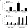
Figure 5. AMJ induced selective pro-apoptotic effects in different human leukemia cells and human primary lymphoblastic leukemia cells.
(A) Pro-apoptotic effect of AMJ was assessed on various human leukemia cells (HSB-2, Molt-4 and CCRF-CEM). Values are shown as means ± S.E.M. (n = 3); *, P<0.05 versus respective control; (B) The apoptosis rate of untreated or AMJ-treated primary T-lymphocytes from 3 healthy subjects or lymphoblastic leukemia cells from 3 patients was assessed by flow cytometry using the annexin V-FITC/PI apoptosis assay. The bar graph shows the cumulative percentage of cells which are in apoptosis.

References

    1. Surh YJ. Cancer chemoprevention with dietary phytochemicals. Nat Rev Cancer. 2003;3:768–780. - PubMed
    1. Chung FL, Schwartz J, Herzog CR, Yang YM. Tea and cancer prevention: studies in animals and humans. J Nutr. 2003;133:3268S–3274S. - PubMed
    1. Sharif T, Auger C, Alhosin M, Ebel C, Achour M, et al. Red wine polyphenols cause growth inhibition and apoptosis in acute lymphoblastic leukaemia cells by inducing a redox-sensitive up-regulation of p73 and down-regulation of UHRF1. Eur J Cancer. 2010;46:983–994. - PubMed
    1. Walter A, Etienne-Selloum N, Brasse D, Khallouf H, Bronner C, et al. Intake of grape-derived polyphenols reduces C26 tumor growth by inhibiting angiogenesis and inducing apoptosis. FASEB J. 2010;24:3360–3369. - PubMed
    1. Hakimuddin F, Paliyath G, Meckling K. Treatment of mcf-7 breast cancer cells with a red grape wine polyphenol fraction results in disruption of calcium homeostasis and cell cycle arrest causing selective cytotoxicity. J Agric Food Chem. 2006;54:7912–7923. - PubMed

Publication types

MeSH terms