. 2012;7(3):e32728.
doi: 10.1371/journal.pone.0032728. Epub 2012 Mar 7.

Commensal bacteria modulate innate immune responses of vaginal epithelial cell multilayer cultures

Affiliations

Commensal bacteria modulate innate immune responses of vaginal epithelial cell multilayer cultures

William A Rose 2nd et al. PLoS One. 2012.

Abstract

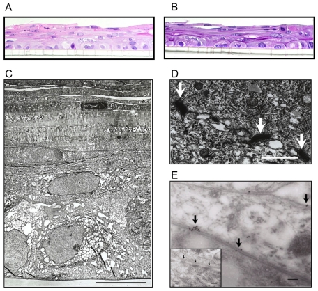
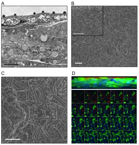
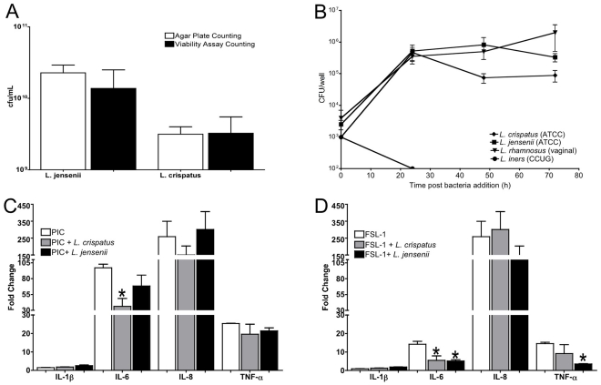
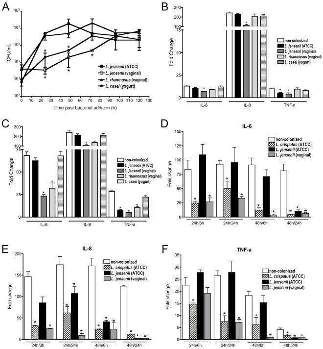
The human vaginal microbiome plays a critical but poorly defined role in reproductive health. Vaginal microbiome alterations are associated with increased susceptibility to sexually-transmitted infections (STI) possibly due to related changes in innate defense responses from epithelial cells. Study of the impact of commensal bacteria on the vaginal mucosal surface has been hindered by current vaginal epithelial cell (VEC) culture systems that lack an appropriate interface between the apical surface of stratified squamous epithelium and the air-filled vaginal lumen. Therefore we developed a reproducible multilayer VEC culture system with an apical (luminal) air-interface that supported colonization with selected commensal bacteria. Multilayer VEC developed tight-junctions and other hallmarks of the vaginal mucosa including predictable proinflammatory cytokine secretion following TLR stimulation. Colonization of multilayers by common vaginal commensals including Lactobacillus crispatus, L. jensenii, and L. rhamnosus led to intimate associations with the VEC exclusively on the apical surface. Vaginal commensals did not trigger cytokine secretion but Staphylococcus epidermidis, a skin commensal, was inflammatory. Lactobacilli reduced cytokine secretion in an isolate-specific fashion following TLR stimulation. This tempering of inflammation offers a potential explanation for increased susceptibility to STI in the absence of common commensals and has implications for testing of potential STI preventatives.


Conflict of interest statement

Competing Interests: The authors have declared that no competing interests exist.

Figures

Figure 1
Figure 1. Human immortalized VEC differentiated into multilayers with air-interfaces morphologically similar to vaginal stratified squamous epithelium.
A. A representative H&E (400×) staining of a d 9 V19I VEC multilayer 5 um section that illustrates the formation of multiple cell layers. The transwell porous membrane can be seen at the bottom of the micrograph. B. PAS staining of a 5 uM section through a VEC multilayer illustrated the substantial amounts of glycogen produced. VEC grown in standard format did not stain by PAS (data not shown). C. A representative transmission EM image of a d 9 V19I VEC multilayer culture (>7 layers) including the transwell porous support (bottom) and the apical anucleate layers sloughing from the top surface (bar = 10 uM). The micrograph illustrates the intracellular structures and the formation of tight junctions indicating full polarization and differentiation. D. Tight junction complexes (white arrows) were observed between the plasma membranes of VEC within 7–9 d of plating (bar = 100 nm) and were confirmed, E., by immunogold labeling for occludin (black arrows show gold particle deposition; bar = 100 nM). Inset panel: A secondary antibody control transmission EM showed no labeling at the tight junction complexes (black arrows, bar = 100 nM).
Figure 2
Figure 2. Commensal bacteria colonized the apical surface of air-interface VEC multilayer cultures.
A. A representative transmission EM micrograph showing the top half of a VEC multilayer culture 24 h post colonization with L. jensenii on the apical surface illustrating the glycogen vesicles contained in the VEC that increased in size and density in the apical most layers of the cultures (bar = 2 um; 14,100×). B. Scanning EM micrographs illustrated the apical colonization of L. jensenii (24 h) and the intimate associations between bacteria and VEC microvilli present on the surface (bar = 2 um; 3000×). Apparent daughter bacterial cells with shorter length as well as areas of higher bacterial density were observed (inset panel). C. Commensal bacteria did not associate with the surfaces of cells that were condensed and apparently sloughing as indicated by their position above the average plane of the apical surface (e.g. cell left side of image; bar = 2 um; 10,000×). D. To confirm the location of the Lactobacilli colonizing the VEC multilayers, confocal Z-axis projection (63×; top panel) and optical slices (bottom panels in order) from a GFP-expressing VEC multilayer (green) with DAPI stained nuclei (blue) and antibody labeled L. jensenii (red) illustrated exclusive superficial colonization of the apical cell surface. No evidence of bacteria (red labeling) in sections below the apical surface was obtained even after 72 h of colonization.
Figure 3
Figure 3. Commensal bacteria colonization kinetics and impact on VEC multilayer cultures.
A. A higher-throughput broth-based bacterial titration system was validated against standard agar plating and subsequent colony counting. Comparison of the two methods showed no statistical differences in viability quantification (p>0.05, Student's t-test) supporting subsequent use of the broth-based viability assay. Data are the mean ± SEM of duplicate samples from 2 independent experiments. B. Replication kinetics and steady state formation of L. crispatus, L. jensenii or L. iners (103 cfu/well) in VEC multilayer cultures are shown over 72 h. L. iners viability rapidly decreased and no viable bacteria were observed after 24 h. L. crispatus and L. jensenii viability significantly (*, p<0.05, Student's t-test) increased after 24 h of culture and was maintained over the study period. Each time point for the selected bacterial species represents the mean ± SEM for triplicate wells of each VEC type (V11I, V12I, V19I) from 2 independent experiments. C and D. Colonization of the VEC by L. crispatus and L. jensenii differently impacted responses to TLR agonists (PIC or FSL-1, respectively) as indicated by the fold change in cytokine levels relative to non-colonized cultures treated in parallel. Data are the mean ± SEM of 3 replicates from a representative experiment out of 3 independent experiments.
Figure 4
Figure 4. Commensal bacteria colonization from low pass clinical isolates and lab adapted strains showed distinct alterations in TLR agonist-induced cytokine expression.
A. Colonization kinetics for several vaginal and non-vaginal Lactobacilli are presented over 5 d with statistically delayed growth for several of the isolates relative to the L. jensenii ATCC type strain (*p<0.05, ANOVA) providing the opportunity to evaluate the impact of bacterial density on cytokine induction. B and C. Data from the standard paradigm indicated that after 24 h of bacterial colonization and 6 h of TLR agonist exposure (FSL-1 or PIC, panels B and C, respectively) showed even reduced titers of the indicated Lactobacilli strains could significantly reduce cytokine induction relative to non-colonized controls treated in parallel (*p<0.05, ANOVA). Colonization with the clinical L. jensenii (vaginal) isolate that reached 24 h titers ∼100-fold lower than the lab adapted isolate (ATCC) produced the most robust reduction in cytokine expression with more pronounced effects following PIC exposure (panel C). D–F. Given the different kinetics of colonization observed for each isolate we also kinetically tested selected bacteria for impact on PIC cytokine induction following either 24 or 48 h of colonization. TLR agonist exposures also were evaluated at two time points (6 and 24 h) creating four testing conditions both confirming and extending the previous observations. The data are presented as fold change relative to non-colonized cultures treated with PIC in parallel for IL-6 (D), IL-8 (E) and TNFa (F; * p<0.05).

References

    1. Eschenbach DA, Thwin SS, Patton DL, Hooton TM, Stapleton AE, et al. Influence of the normal menstrual cycle on vaginal tissue, discharge, and microflora. Clin Infect Dis. 2000;30:901–907. - PubMed
    1. Farage M, Maibach H. Lifetime changes in the vulva and vagina. Arch Gynecol Obstet. 2006;273:195–202. - PubMed
    1. Hladik F, Sakchalathorn P, Ballweber L, Lentz G, Fialkow M, et al. Initial events in establishing vaginal entry and infection by human immunodeficiency virus type-1. Immunity. 2007;26:257–270. - PMC - PubMed
    1. Farage MA, Maibach HI. Morphology and physiological changes of genital skin and mucosa. Curr Probl Dermatol. 2011;40:9–19. - PubMed
    1. Ayehunie S, Cannon C, Lamore S, Kubilus J, Anderson DJ, et al. Organotypic human vaginal-ectocervical tissue model for irritation studies of spermicides, microbicides, and feminine-care products. Toxicol In Vitro. 2006;20:689–698. - PubMed

Publication types

MeSH terms

LinkOut - more resources