. 2012;7(3):e32777.
doi: 10.1371/journal.pone.0032777. Epub 2012 Mar 7.

Evolution of JAK-STAT pathway components: mechanisms and role in immune system development

Affiliations

Evolution of JAK-STAT pathway components: mechanisms and role in immune system development

Clifford Liongue et al. PLoS One. 2012.

Abstract

Background: Lying downstream of a myriad of cytokine receptors, the Janus kinase (JAK)-Signal transducer and activator of transcription (STAT) pathway is pivotal for the development and function of the immune system, with additional important roles in other biological systems. To gain further insight into immune system evolution, we have performed a comprehensive bioinformatic analysis of the JAK-STAT pathway components, including the key negative regulators of this pathway, the SH2-domain containing tyrosine phosphatase (SHP), Protein inhibitors against Stats (PIAS), and Suppressor of cytokine signaling (SOCS) proteins across a diverse range of organisms.

Results: Our analysis has demonstrated significant expansion of JAK-STAT pathway components co-incident with the emergence of adaptive immunity, with whole genome duplication being the principal mechanism for generating this additional diversity. In contrast, expansion of upstream cytokine receptors appears to be a pivotal driver for the differential diversification of specific pathway components.

Conclusion: Diversification of JAK-STAT pathway components during early vertebrate development occurred concurrently with a major expansion of upstream cytokine receptors and two rounds of whole genome duplications. This produced an intricate cell-cell communication system that has made a significant contribution to the evolution of the immune system, particularly the emergence of adaptive immunity.


Conflict of interest statement

Competing Interests: The authors have declared that no competing interests exist.

Figures

Figure 1
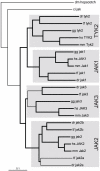
Figure 1. Phylogenetic analysis of JAK family members.
Phylogenetic analysis of JAK protein sequences using the Neighborhood-Joining algorithm, with bootstrap values above 80% (of 1000 replicates) indicated in bold. Species used for phylogenetic analysis are indicated: fruit fly (dm), sea squirt (ci), zebrafish (dr), green pufferfish (tf), Japanese pufferfish (tr), chicken (gg), mouse (mm), and human (hs).
Figure 2
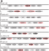
Figure 2. Synteny analysis of JAK family members.
Synteny analysis of the relevant zebrafish and human JAK gene loci (white), indicating adjacent genes in their respective orientations. Genes showing conserved synteny between zebrafish and human are in black, genes which have homologues displaying synteny with other family members in humans or zebrafish edged in red and non-syntenic genes in grey.
Figure 3
Figure 3. Phylogenetic analysis of STAT family members.
Phylogenetic analysis of STAT family members as described for Figure 1, with the inclusion of genes from African clawed frog (xl) and Western clawed frog (xt).
Figure 4
Figure 4. Synteny analysis of STAT family members.
Conserved synteny analysis of STAT loci as described in Figure 2, with the addition of genes that displayed synteny between zebrafish and Japanese pufferfish being shown in green.
Figure 5
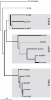
Figure 5. Phylogenetic analysis of SHP family members.
Phylogenetic analysis of SHP family members as described for Figure 3.
Figure 6
Figure 6. Synteny analysis of SHP family members.
Syntenic analysis of SHP family members as described for Figure 4.
Figure 7
Figure 7. Phylogenetic analysis of PIAS family members.
Phylogenetic analysis of PIAS family members as described for Figure 3.
Figure 8
Figure 8. Synteny analysis of PIAS family members.
Syntenic analysis of PIAS family members as described for Figure 4.
Figure 9
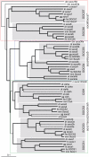
Figure 9. Phylogenetic analysis of SOCS family members.
Phylogenetic analysis was performed on SOCS family members as described for Figure 3.
Figure 10
Figure 10. Synteny analysis of SOCS family members.
Syntenic analysis of SOCS family members as described for Figure 4.
Figure 11
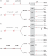
Figure 11. A model for the evolution of the of JAK, SHP, and PIAS families.
(A) Evolutionary timeline summarizing key events in chordate/vertebrate evolution. Key evolutionary events are shown, including the divergence of protostomes (solid grey line) from deuterostomes, the subsequent divergence of urochordates (broken grey line) from other chordates, and finally ray-finned fish (including teleosts) (dotted black line) from lobe-finned fish (including terapods) (black line), with relevant present-day groupings (insect, urochordate, teleost, and tetrapod) indicated. Whole genome duplications (1R, 2R, 3R) are indicated with short red lines. (B–D) Model for the evolution of JAK-STAT pathway components: JAK (B), SHP (C), and PIAS (D) families. Red ovals indicate the likely core members at the time of protostomes/deuterostomes and urochordate/chordate divergence, while the grey shaded rectangle represents ray-finned/lobe-finned fish ‘core’ signaling components present at the time of divergence. Lineage-specific/local duplications are indicated by red dots and gene conversion indicated by a red square.
Figure 12
Figure 12. A model for the evolution of the STAT family.
A model for the evolution of the STAT component of the JAK-STAT pathway as described in Figure 11.
Figure 13
Figure 13. A model for the evolution of the SOCS family.
A model for the evolution of the SOCS component of the JAK-STAT pathway as described in Figure 11.

References

    1. Ozaki K, Leonard WJ. Cytokine and cytokine receptor pleiotropy and redundancy. J Biol Chem. 2002;277:29355–29358. - PubMed
    1. Voss SD, Hong R, Sondel PM. Severe combined immunodeficiency, interleukin-2 (IL-2), and the IL-2 receptor: experiments of nature continue to point the way. Blood. 1994;83:626–635. - PubMed
    1. Kotenko SV, Gallagher G, Baurin VV, Lewis-Antes A, Shen M, et al. IFN-lambdas mediate antiviral protection through a distinct class II cytokine receptor complex. Nat Immunol. 2003;4:69–77. - PubMed
    1. Liongue C, Wright C, Russell AP, Ward AC. Granulocyte colony-stimulating factor receptor: stimulating granulopoiesis and much more. Int J Biochem Cell Biol. 2009;41:2372–2375. - PubMed
    1. Seubert N, Royer Y, Staerk J, Kubatzky KF, Moucadel V, et al. Active and inactive orientations of the transmembrane and cytosolic domains of the erythropoietin receptor dimer. Mol Cell. 2003;12:1239–1250. - PubMed

Publication types

MeSH terms

Substances