. 2012;7(3):e33289.
doi: 10.1371/journal.pone.0033289. Epub 2012 Mar 7.

Cadherin-23 mediates heterotypic cell-cell adhesion between breast cancer epithelial cells and fibroblasts

Affiliations

Cadherin-23 mediates heterotypic cell-cell adhesion between breast cancer epithelial cells and fibroblasts

Maria Apostolopoulou et al. PLoS One. 2012.

Abstract

In the early stages of breast cancer metastasis, epithelial cells penetrate the basement membrane and invade the surrounding stroma, where they encounter fibroblasts. Paracrine signaling between fibroblasts and epithelial tumor cells contributes to the metastatic cascade, but little is known about the role of adhesive contacts between these two cell types in metastasis. Here we show that MCF-7 breast cancer epithelial cells and normal breast fibroblasts form heterotypic adhesions when grown together in co-culture, as evidenced by adhesion assays. PCR and immunoblotting show that both cell types express multiple members of the cadherin superfamily, including the atypical cadherin, cadherin-23, when grown in isolation and in co-culture. Immunocytochemistry experiments show that cadherin-23 localizes to homotypic adhesions between MCF-7 cells and also to heterotypic adhesions between the epithelial cells and fibroblasts, and antibody inhibition and RNAi experiments show that cadherin-23 plays a role in mediating these adhesive interactions. Finally, we show that cadherin-23 is upregulated in breast cancer tissue samples, and we hypothesize that heterotypic adhesions mediated by this atypical cadherin may play a role in the early stages of metastasis.


Conflict of interest statement

Competing Interests: The authors have declared that no competing interests exist.

Figures

Figure 1
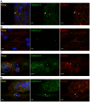
Figure 1. MCF-7s and NBFs form heterotypic adhesions and express cadherin-23 in vitro.
A) NBFs were pre-labeled with CellTracker™ Green and co-cultured with MCF-7s for 3 days. Cells were then fixed and labeled with an antibody against cytokeratin-8 (red). Arrow shows a site of heterotypic contact between the two cell types. B) Three-day co-cultures labeled with antibodies against β-catenin (red) and cytokeratin-8 (green). Arrow – heterotypic contact; Arrowhead – MCF-7 homotypic contact. Scale = 10 µm. C) MCF-7s labeled with CellTracker Green™ (arrowhead) were co-cultured with NBFs labeled with CellTracker Red™ (arrow). Phase contrast (left) and fluorescence image (right) of a typical heterotypic cell aggregate are shown. Scale = 20 µm. D) Immunoblots show no vimentin expression in MCF-7 fraction and no cytokeratin-8 in NBF fraction after FACS separation of co-cultures. PCR (E) and immunoblots (F) showing cadherin-23 expression in MCF-7s and NBFs before and after co-culture. α-tubulin and GAPDH were used as controls.
Figure 2
Figure 2. Cadherin-23 localizes to homotypic and heterotypic contacts.
Cadherin-23 localizes at 75% of homotypic contacts between MCF-7s (arrowhead, A) but not NBFs (B). Arrow in (A) points to a contact between MCF-7s without cadherin-23 recruitment. Cadherin-23 localizes at 23% of heterotypic adhesions in two-hour (C) and 45% of three-day (D) co-cultures (arrowhead). Asterisks in (C–D) mark fibroblasts. Fig. 2C shows MCF-7 cells and NBFs co-cultured for two hours, where a group of spherical MCF-7 cells is on top of a flat and extended fibroblast (asterisk). One of the MCF-7 cells has formed a possible adhesion site with the fibroblast, as evidenced by β-catenin labeling, and cadherin-23 has already been recruited at that site (arrowhead). Fig. 2D shows a group of compact MCF-7 cells, where one of them has formed an adhesion site with an extended fibroblast (asterisk), as marked by β-catenin localization, where cadherin-23 has been recruited (arrowhead). All images are maximum projections of confocal z-stacks. Scale = 10 µm.
Figure 3
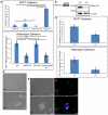
Figure 3. Cadherin-23 plays a role in homotypic and heterotypic adhesion.
A) Adhesion assays using inhibitory antibodies. Top panel: the percent of total MCF-7 cell aggregates comprised of 3 or more cells was measured at t = 0 and at t = 2 hrs and the change in this percentage was calculated. Cadherin-23 and E-cadherin antibodies, individually and in conjunction, showed a statistically significant reduction of adhesion when compared to the effect of an antibody that blocks OB-cadherin-mediated adhesion. (n = 3 independent experiments; one tail unpaired t-test; asterisks mark statistically significant differences). Bottom panel: adhesion assays on 1∶1 mixture of MCF-7:NBF after 2 hrs. The antibody against cadherin-23, both alone and in conjunction with the E-cadherin antibody, significantly reduced heterotypic adhesion. (n = 6 independent experiments; one tail unpaired t-test; asterisks mark statistically significant differences; NS = not significant). B) RNAi knock down of cadherin-23 in MCF-7 cells, as evidenced by both RT-PCR (left panel) and immunoblotting (right panel). In the left panel, 25 ng of mRNA were used for each PCR reaction, and the entire product was run on the agarose gel. NC: non-coding RNAi sequence; KD: cadherin-23 specific RNAi sequence. C) Adhesion assays, as in panel 3A, but after RNAi knock down of cadherin-23 in MCF-7 cells. Top panel: RNAi treatment significantly reduced the ability of MCF-7 cells to participate in homotypic adhesion. (n = 3; one tail unpaired t-test; asterisks mark statistically significant differences). Bottom panel: RNAi treatment significantly reduced the number of heterotypic aggregates after 2 hrs. (n = 3; one tail unpaired t-test; asterisks mark statistically significant differences). D) Representative image of MCF-7 cell aggregates after treatment with RNAi. Scale = 100 µm. E) Representative image of MCF-7:NBF cell aggregates after treatment with RNAi. shRNA plasmids contained a gene to a green fluorescent protein, while NBFs were labeled with CellTracker™ Red. Hoescht dye was used to stain the nuclei.
Figure 4
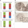
Figure 4. Cadherin-23 is upregulated in breast cancer.
Samples from breast cancer (n = 58, including both in situ and invasive carcinomas) and healthy breast tissue (n = 8) were labeled with cadherin-23 antibodies and blindly scored for overall (top panel), stromal (middle panel) and epithelial (bottom panel) cadherin-23 labeling (‘−’: no expression; ‘+’: low expression; ‘++’: high expression) (A). In all cases carcinoma samples showed significantly higher expression (Wilcoxon Rank-Sum test, p = 0.01). Representative carcinoma (in situ) (B) (scored as ‘++’) and normal breast (C) tissue (scored as ‘−’) labeled with cadherin-23 antibodies. Arrow in (B) points to a duct with intense cadherin-23 labeling that is budding into the stroma. Insets in (B–C) show a higher magnification of the enclosed area. Scale = 0.1 mm.

References

    1. Samoszuk M, Tan J, Chorn G. Clonogenic growth of human breast cancer cells co-cultured in direct contact with serum-activated fibroblasts. Breast Cancer Res. 2005;7:R274–283. - PMC - PubMed
    1. Omelchenko T, Fetisova E, Ivanova O, Bonder EM, Feder H, et al. Contact interactions between epitheliocytes and fibroblasts: formation of heterotypic cadherin-containing adhesion sites is accompanied by local cytoskeletal reorgnization. PNAS. 2001;98:8632–8637. - PMC - PubMed
    1. Stemmler MP. Cadherins in development and cancer. Mol Biosyst. 2008;4:835–850. - PubMed
    1. Nieman MT, Prudoff RS, Johnson KR, Wheelock MJ. N-cadherin promotes motility in human breast cancer cells regardless of their E-cadherin expression. J Cell Biol. 1999;147:631–644. - PMC - PubMed
    1. Feltes CM, Kudo A, Blaschuk O, Byers SW. An alternatively spliced cadherin-11 enhances human breast cancer cell invasion. Cancer Res. 2002;62:6688–6697. - PubMed

Publication types