. 2012 Apr 3;84(7):3170-8.
doi: 10.1021/ac203112c. Epub 2012 Mar 20.

Combined elemental and biomolecular mass spectrometry imaging for probing the inventory of tissue at a micrometer scale

Affiliations

Combined elemental and biomolecular mass spectrometry imaging for probing the inventory of tissue at a micrometer scale

Andreas Matusch et al. Anal Chem. .

Abstract

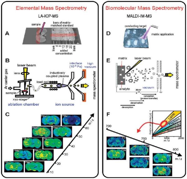
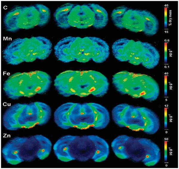
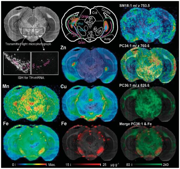
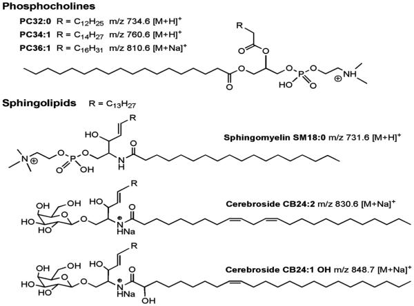
Several complementary mass spectrometric imaging techniques allow mapping of various analytes within biological tissue sections. Laser ablation inductively coupled plasma mass spectrometry (LA-ICPMS) quantitatively detects elements and isotopes with very high sensitivity and a particularly high dynamical range. Matrix-assisted laser desorption/ionization ion mobility mass spectrometry (MALDI-IM-MS) allows a pixel-by-pixel classification and identification of biomolecules. In order to dispose of the healthy hemisphere as an internal calibrant in addition to routinely used external standards, adjacent brain sections of mice with a unilateral 6-OHDA lesion of the medial forebrain bundle were chosen as exemplary samples. We demonstrate a comprehensive way of data acquisition and analysis by coregistering mass spectrometric data on photomicrographs as common reference space and thus providing trimodal spatial information. Registering subsequent planar element maps yielded continuous 3-dimensional data sets. Furthermore, we introduce a correction of MSI data for variable slice thickness applicable to all MSI techniques. In the present case, we observed increased concentrations of iron, manganese, and copper in the lesioned substantia nigra while monounsaturated lipid levels were decreased in the identical region of interest. Our techniques provide new insights into the intricate spatial relationship of morphology and chemistry within tissue.


Figures

Figure 1
Figure 1
Workflow of LA-ICPMS imaging (A–C) and MALDI-IMMS imaging (D–F): Cryosections were placed together with a ladder of matrix matched standards on a glass slide (A) and on a conductive MALDI plate and DHB was applied as matrix onto the samples (D). B and E show the processes preceding ion separation which is laser ablation followed by inductively coupled plasma ionization (B) or the sequence of laser-induced desorption, desolvation, and ionization within the MALDI source. Exemplary resulting ion maps on a m/z scale are given in (C) and (F). The inset in (F) shows schematically the position of molecule classes in IM-MS conformational space constituted by m/z and the drift time reflecting the collision cross section. Ion images selectively corresponding to lipids are displayed.
Figure 2
Figure 2
Three-dimensional reconstructions obtained by affine registration of 11 LA-ICPMS elemental concentration maps of adjacent coronal sections through the substantia nigra (SN). Three semitransparent maximum intensity projections from different angels of five elements are shown. The SN at the lesioned (right) side clearly shows higher iron than the contralateral healthy SN. The width of one section is 9 mm.
Figure 3
Figure 3
Multimodal imaging approach integrating light microscopy underlain as background of each image facilitating morphological orientation, elemental concentration maps obtained by LA-ICPMS (left and middle column) and lipid maps obtained by MALDI-IM-MS imaging. The respective maximum concentrations of the color scale are Zn, 50; Mn, 0.6 Cu, 10 and Fe, 35 μg g−1. The lipid maps shown represent the tree major distribution types observed, cortical (m/z 753.5 assigned to sphingomyelin SM18:1), cortico-mesencephalic (m/z 760.6 assigned to phosphatidylcholine PC34:1), and of white matter predominance (m/z 826.6 assigned to phosphatidylcholine PC36:1). The picture at the bottom right shows an overlay of light microscopy, LA-ICPMS image (Fe) and MALDI-IM-MS image. There is an increase of Fe and a decrease of the monounsaturated lipid within the right substantia nigra (SN). Darkfield images across the entire SN of sections subjacent to sections used for MS demonstrate ISH signals of TH-positive dopaminergic neurons. Note the nearly complete loss of dopaminergic neurons in the SN pars compacta and ventral tegmental area in the lesioned right side compared to control left side. The schema in the middle of the upper row explains the corresponding anatomy. Abbreviations: Amy, amygdala; Aq, aqueduct; c, corpus callosum (composed of white matter); Col, colliculus superior; Gran, granular layer of the fascia dentata (cut at two positions, dorsal and a ventral); Hip, hippocampus (green dashed conture); if, interfascicular nucleus; ip, interpeduncular region; Mol, stratum moleculare of the fascia dentate; o, accessory oculomotor nuclei; PAG, periaqueductal gray; R, nucleus ruber; Pol, polymorph layer (= stratum multiforme) of the fascia dentata; SLu, stratum lucidum of cornu ammonis portion 3 (of CA3); SN, substantia nigra.
Figure 4
Figure 4
Structures of selected lipids and ion species detected by MALDI-IM-MS.

References

    1. Chughtai K, Heeren RMA. Chem. Rev. 2010;110:3237–3277. - PMC - PubMed
    1. Rubakhin SS, Sweedler J. V. e. Mass Spectrometric Imaging: Principles and Protocols. Vol. 656. Springer; Heidelberg: 2010.
    1. Altelaar AFM, Luxembourg SL, McDonnell LA, Piersma SR, Heeren RMA. Nat. Protoc. 2007;2:1185–1196. - PubMed
    1. Stoeckli M, Chaurand P, Hallahan DE, Caprioli RM. Nat. Med. 2001;7:493–496. - PubMed
    1. Caprioli RM, Farmer TB, Gile J. Anal. Chem. 1997;69:4751–4760. - PubMed

Publication types