. 2012 Jun 15;7(6):1067-76.
doi: 10.1021/cb200525q. Epub 2012 Apr 13.

Targeting native adult heart progenitors with cardiogenic small molecules

Affiliations

Targeting native adult heart progenitors with cardiogenic small molecules

Jamie L Russell et al. ACS Chem Biol. .

Abstract

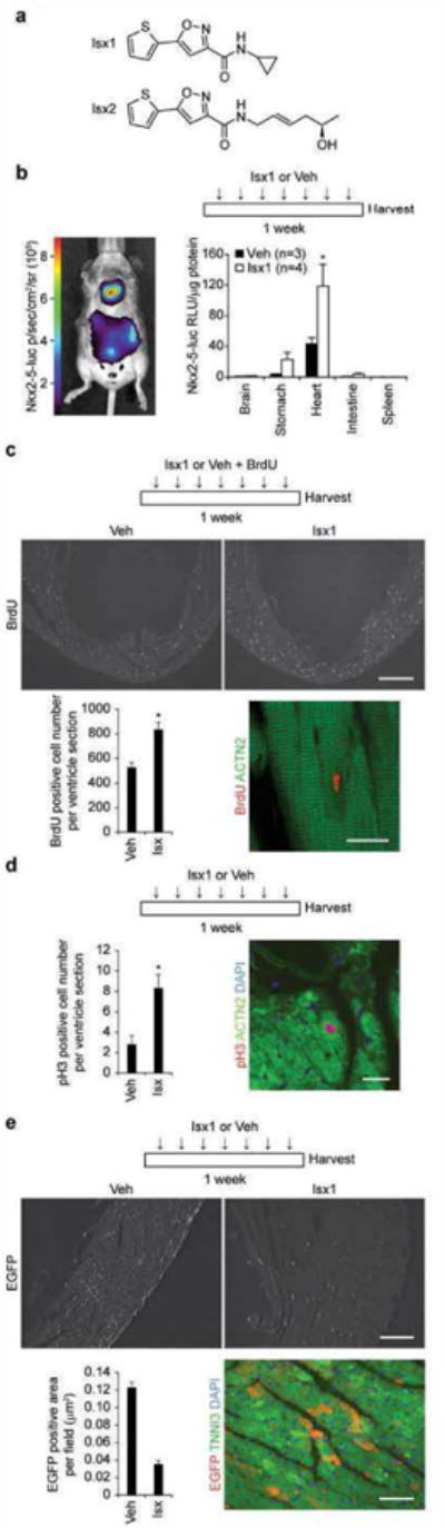
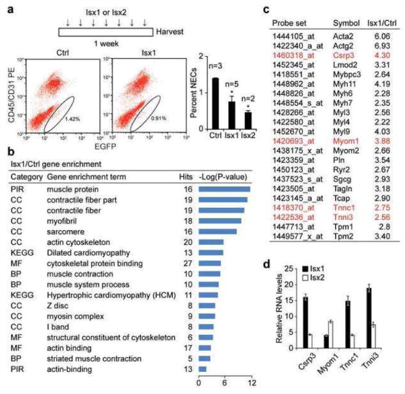
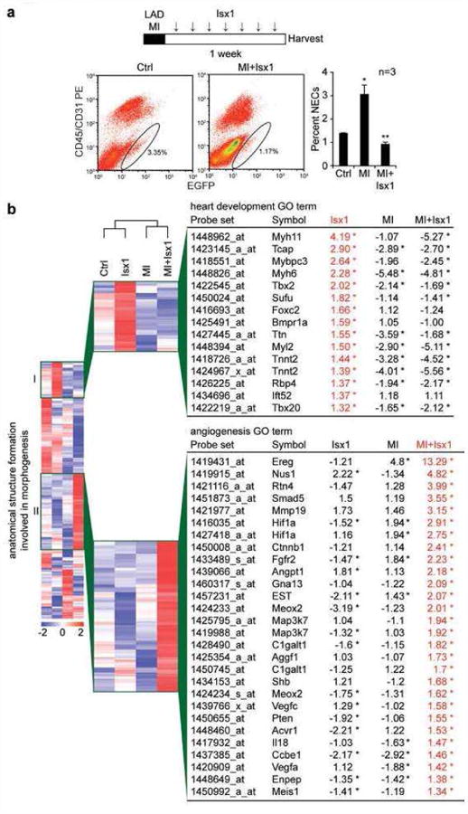
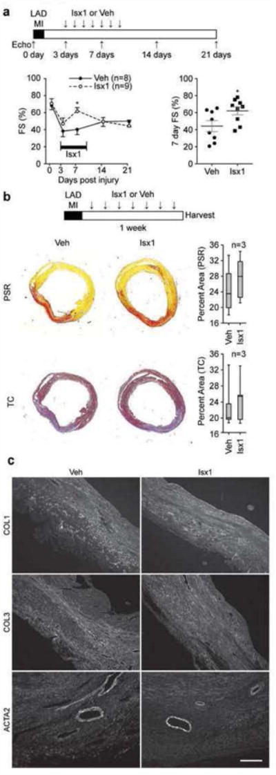
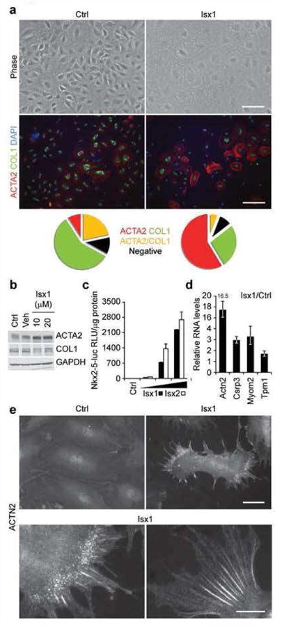
Targeting native progenitors with small molecule pharmaceuticals that direct cell fate decisions is an attractive approach for regenerative medicine. Here, we show that 3,5-disubstituted isoxazoles (Isx), stem cell-modulator small molecules originally recovered in a P19 embryonal carcinoma cell-based screen, directed cardiac muscle gene expression in vivo in target tissues of adult transgenic reporter mice. Isx also stimulated adult mouse myocardial cell cycle activity. Narrowing our focus onto one target cardiac-resident progenitor population, Isx directed muscle transcriptional programs in vivo in multipotent Notch-activated epicardium-derived cells (NECs), generating Notch-activated adult cardiomyocyte-like precursors. Myocardial infarction (MI) preemptively differentiated NECs toward fibroblast lineages, overriding Isx's cardiogenic influence in this cell population. Isx dysregulated gene expression in vivo in Notch-activated repair fibroblasts, driving distinctive (pro-angiogenesis) gene programs, but failed to mitigate fibrosis or avert ventricular functional decline after MI. In NECs in vitro, Isx directed partial muscle differentiation, which included biosynthesis and assembly of sarcomeric α-actinin premyofibrils, beaded structures pathognomonic of early developing cardiomyocytes. Thus, although Isx small molecules have promising in vivo efficacy at the level of cardiac muscle gene expression in native multipotent progenitors and are first in class in this regard, a greater understanding of the dynamic interplay between fibrosis and cardiogenic small molecule signals will be required to pharmacologically enable regenerative repair of the heart.


Figures

Figure 1
Figure 1. Isx1 pharmacologically targets and regulates myocardial cells in vivo
(a) Chemical structures of Isx1 (N-cyclopropyl-5-(thiophen-2-yl)isoxazole-3-carboxamide) and Isx2 ((R,E)-N-(5-hydroxyhex-2-enyl)-5-(thiophen-2-yl)isoxazole-3-carboxamide) compounds. (b) Time map of Isx1 in vivo treatment paradigm. Bioluminescence imaging of Nkx2-5-luc-BAC mouse demonstrating cardiac and stomach (a known extra-cardiac target) restricted luciferase activity after Isx injection in vivo. Quantitative evaluation of Isx-induced Nkx2-5-luc-BAC activity normalized per μg protein in tissue extracts from reporter mice injected ip once daily for 7 days with cyclodextrin (Veh, n=3) or Isx1 at 16 mg/kg (n=4). (c) Time map of Isx1 or Veh plus BrdU in vivo treatment paradigm. BrdU immunohistochemistry in uninjured adult mouse heart injected with vehicle or Isx (top panels, scale bar = 500μm). Quantitative evaluation of BrdU+ cells per ventricle section (9 sections from 3 animals were counted for each group, * = p<0.005). Immunohistochemistry localizing a BrdU+ nucleus (red) to an ACTN2+ cardiomyocyte (green) in an uninjured adult mouse injected with Isx (bottom right panel, Scale bar = 20μm). (d) Time map of Isx1 in vivo treatment paradigm. Quantitative evaluation of pH3+ cells per ventricle section in uninjured adult mice injected with Veh or Isx1 for 7 days (6 sections from 3 animals were counted for each group, * = p<0.05). Phospho-histone H3+ nucleus (red) localizing to an ACTN2+ cardiomyocyte (green), co-stained with DAPI (blue), in an uninjured adult mouse injected with Isx (right hand panel, Scale bar = 30μm). (e) Time map of Isx1 in vivo treatment paradigm. EGFP immunohistochemistry in uninjured adult mouse heart injected with Veh or Isx1 (top panels, scale bar = 100μm). Quantitative evaluation of EGFP+ area per field (18 fields per group) in uninjured adult mice treated with Veh or Isx1 for 7 days. EGFP+ (red) immunohistochemistry localized to TNNI3+ (green) cardiomyocytes, co-stained with DAPI (blue) (bottom right panel, scale bar = 30μm).
Figure 2
Figure 2. Isx chemically programs cardiomyogenic gene expression in NECs in vivo
(a) Time map of Isx1, Isx2 or Veh in vivo treatment paradigm. FACS profiles and graph demonstrating dynamic regulation of NEC (Notch/CBF1-REx4-EGFP+CD45CD31 cell) number per adult age-matched male TNR mouse hearts, after treating mice with Isx1 or Isx2 (n = 3 for Ctrl; n = 5 for Isx1; n = 2 for Isx2; * = p<0.05 comparing NECs from experimental groups to Ctrl; using a two-tailed unpaired Student's t-test). (b) Microarray gene enrichment analysis of NECs from Isx1 versus Ctrl TNR mice demonstrating significant enrichment of cardiac muscle terms in NECs from Isx1 treated mice. (c) A representative list of genes from the gene ontology groups in (b), comparing relative transcript levels in NECs from Isx1 versus Ctrl TNR hearts. (d) Independent confirmation of microarray cardiac gene activation (genes highlighted in red in (c)) by Isx1 in NECs in vivo compared with Isx2 by QPCR.
Figure 3
Figure 3. MI switches NEC's Isx-transcriptional response from cardiomyogenic to fibrosis and angiogenesis genes
(a) Time map of LAD-MI and Isx1 in vivo treatment paradigm. FACS profiles and graph demonstrating dynamic regulation of NEC (Notch/CBF1-REx4-EGFP+CD45CD31 cell) number in adult age-matched male TNR mouse hearts after LAD-MI and weeklong treatment with Isx1 (n=3 for Ctrl, MI, and MI+Isx1; *=p<0.05 comparing NECs from experimental groups to Ctrl; ** = p<0.01 comparing NECs from MI+Isx1 to MI alone using a two-tailed unpaired Student's t-test). (b) Gene enrichment analysis of Affymetrix microarray data comparing gene expression in NECs isolated from Ctrl, Isx1, MI and MI+Isx1 treated TNR mice. Heat map of the “anatomical structure formation involved in morphogenesis” gene ontology group highlighting an enlarged section of a unique gene cluster with high expression after Isx1 treatment and a list of genes in the “heart development” ontology group (cluster I, top) and a second unique gene cluster with high expression after MI+Isx1 treatment and a list of genes in the “angiogenesis” ontology group (cluster II, bottom), identified by enrichment analysis of the cluster, with fold change of transcript levels for Isx1, MI and MI+Isx1 versus Ctrl (red=high expression; blue=low expression; the asterisk indicates transcripts that are “present” by Affymetrix chip criteria in Ctrl and the indicated samples, and whose transcript levels after Isx1, MI or MI+Isx1 differed from Ctrl, up or down, in a statistically significant manner (see M & M for details).
Figure 4
Figure 4. Isx improves post-MI ventricular function at week one without gross structural changes
(a) Time map of LAD-MI, Isx1 or Veh in vivo treatment and echocardiography paradigm. Isx1 significantly improved ventricular function (fractional shortening) evaluated on post-MI day 7 during Isx1 treatment (time course graph on the left and day 7 post-MI scatterplot on the right; n=8 mice for Veh; n=9 mice for Isx1; * = p<0.05 comparing Isx1 and Ctrl using a two-tailed unpaired Student's t test). (b) Time map of LAD-MI, Isx1 or Veh in vivo treatment paradigm. At week one post-MI, Isx1 treatment did not change scar size (measured by quantitating Picrosirius Red (PSR) or Masson's Trichrome (TC) staining area of myocardial cross-sections) or (c) composition (evaluated by immunohistochemical staining for collagens-1 (COL1) and -3 (COL3) and smooth muscle α–actin (ACTA2) (Scale bar = 200μm).
Figure 5
Figure 5. Isx induces cardiomyogenic differentiation of NECs in vitro
(a) Phase contrast images (top panels, scale bar = 100μm) of clonally-derived NECs cultured for 4 days in media alone (Ctrl) or treated with Isx1 (20 μM), and double-immunostained for ACTA2 (red) and COL1 (green) with DAPI (blue) counter stain (bottom panels, scale bar = 200μm). Pie charts represent the percentage of total cells that were ACTA2+ (red), COL1+ (green), ACTA2+/COL1+ (yellow) or double negative (black). (b) Protein blotting for ACTA2 and COL1 in NECs cultured for 4 days in media alone (Ctrl), in the presence of DMSO (Veh) or increasing amounts of Isx1 with GAPDH to control for loading. (c) Dose-dependent activation of Nkx2-5-luc-BAC transgene in NEC reporter cell line (3G8) by Isx1 and 1sx2. (d) Upregulation of candidate cardiac genes in NECs cultured for 4 days in media alone or in the presence of 20 μM Isx1 by QPCR. (e) ACTN2 immunohistochemistry in NECs cultured for 4 days in media alone (Ctrl-top left panel) or 20 μM Isx1 (top right panel) (Scale bar = 50μm). Bottom panel are higher magnification images of ACTN2 immunohistochemistry in Isx1 treated NECs (Scale bar = 20μm).

References

    1. Tsao L, Gibson CM. Heart failure: an epidemic of the 21st century. Crit Pathw Cardiol. 2004;3:194–204. - PubMed
    1. Daley GQ, Scadden DT. Prospects for stem cell-based therapy. Cell. 2008;132:544–548. - PubMed
    1. Zhou B, Honor LB, He H, Ma Q, Oh JH, Butterfield C, Lin RZ, Melero-Martin JM, Dolmatova E, Duffy HS, Gise A, Zhou P, Hu YW, Wang G, Zhang B, Wang L, Hall JL, Moses MA, McGowan FX, Pu WT. Adult mouse epicardium modulates myocardial injury by secreting paracrine factors. J Clin Invest. 121:1894–1904. - PMC - PubMed
    1. Grayson P, Mendlein J, Thies S, Yingling J. Stem cell modulators (SCMs): a small molecule and biologic approach to stem cell therapeutics. Drug Discovery Today: Therapeutic Strategies. 2009;6:141–145.
    1. Adams GB, Martin RP, Alley IR, Chabner KT, Cohen KS, Calvi LM, Kronenberg HM, Scadden DT. Therapeutic targeting of a stem cell niche. Nature biotechnology. 2007;25:238–243. - PubMed

Publication types

MeSH terms