

3D Variation in delineation of head and neck organs at risk
- PMID: 22414264
- PMCID: PMC3337234
- DOI: 10.1186/1748-717X-7-32
3D Variation in delineation of head and neck organs at risk
Abstract
Background: Consistent delineation of patient anatomy becomes increasingly important with the growing use of highly conformal and adaptive radiotherapy techniques. This study investigates the magnitude and 3D localization of interobserver variability of organs at risk (OARs) in the head and neck area with application of delineation guidelines, to establish measures to reduce current redundant variability in delineation practice.
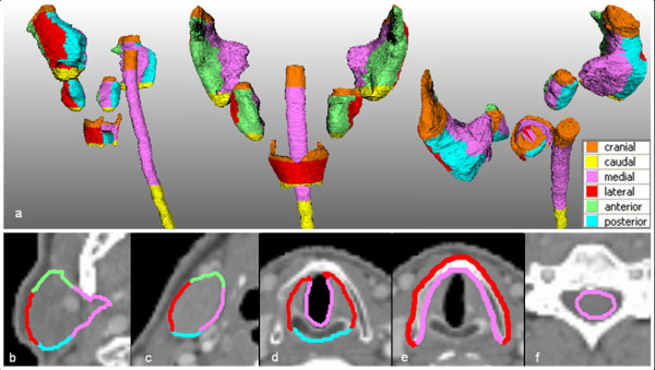
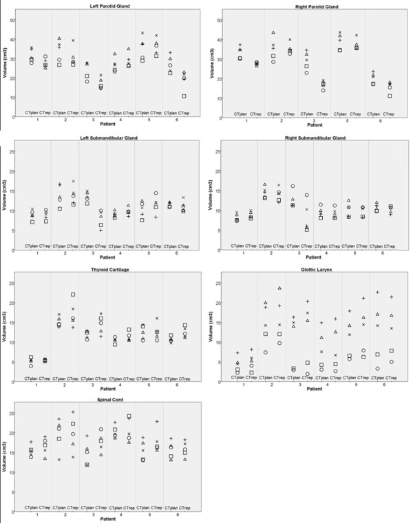
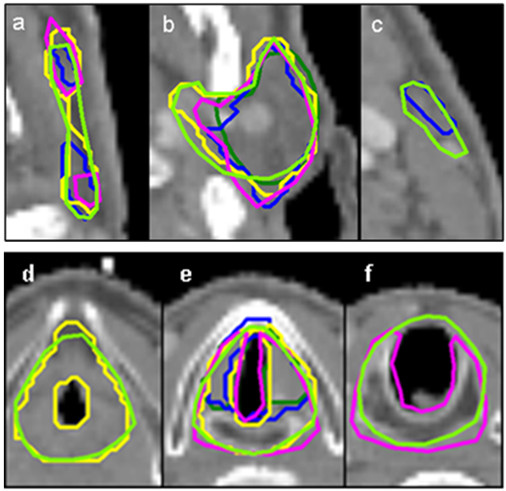
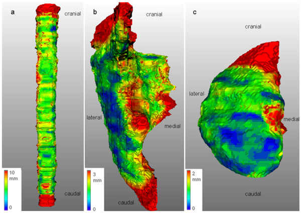
Methods: Interobserver variability among five experienced radiation oncologists was studied in a set of 12 head and neck patient CT scans for the spinal cord, parotid and submandibular glands, thyroid cartilage, and glottic larynx. For all OARs, three endpoints were calculated: the Intraclass Correlation Coefficient (ICC), the Concordance Index (CI) and a 3D measure of variation (3D SD).
Results: All endpoints showed largest interobserver variability for the glottic larynx (ICC = 0.27, mean CI = 0.37 and 3D SD = 3.9 mm). Better agreement in delineations was observed for the other OARs (range, ICC = 0.32-0.83, mean CI = 0.64-0.71 and 3D SD = 0.9-2.6 mm). Cranial, caudal, and medial regions of the OARs showed largest variations. All endpoints provided support for improvement of delineation practice.
Conclusions: Variation in delineation is traced to several regional causes. Measures to reduce this variation can be: (1) guideline development, (2) joint delineation review sessions and (3) application of multimodality imaging. Improvement of delineation practice is needed to standardize patient treatments.
Figures
References
-
- Peters LJ, O'Sullivan B, Giralt J, Fitzgerald TJ, Trotti A, Bernier J, Bourhis J, Yuen K, Fisher R, Rischin D. Critical impact of radiotherapy protocol compliance and quality in the treatment of advanced head and neck cancer: results from TROG 02.02. J Clin Oncol. 2010;28:2996–3001. doi: 10.1200/JCO.2009.27.4498. - DOI - PubMed
-
- Nelms BE, Tomé WA, Robinson G, Wheeler J. Variations in the contouring of organs at risk: test case from a patient with oropharyngeal cancer. Int J Radiat Oncol Biol Phys. in press . - PubMed
MeSH terms
LinkOut - more resources
Full Text Sources
Other Literature Sources
Medical
