Review
. 2012 Aug;69(16):2727-38.
doi: 10.1007/s00018-012-0952-2. Epub 2012 Mar 14.

Interkinetic nuclear migration: beyond a hallmark of neurogenesis

Affiliations
Review

Interkinetic nuclear migration: beyond a hallmark of neurogenesis

Yoichi Kosodo. Cell Mol Life Sci. 2012 Aug.

Abstract

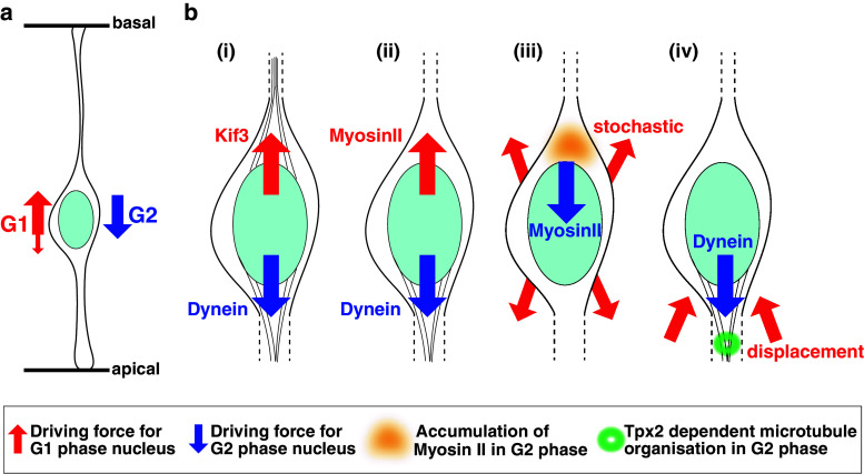
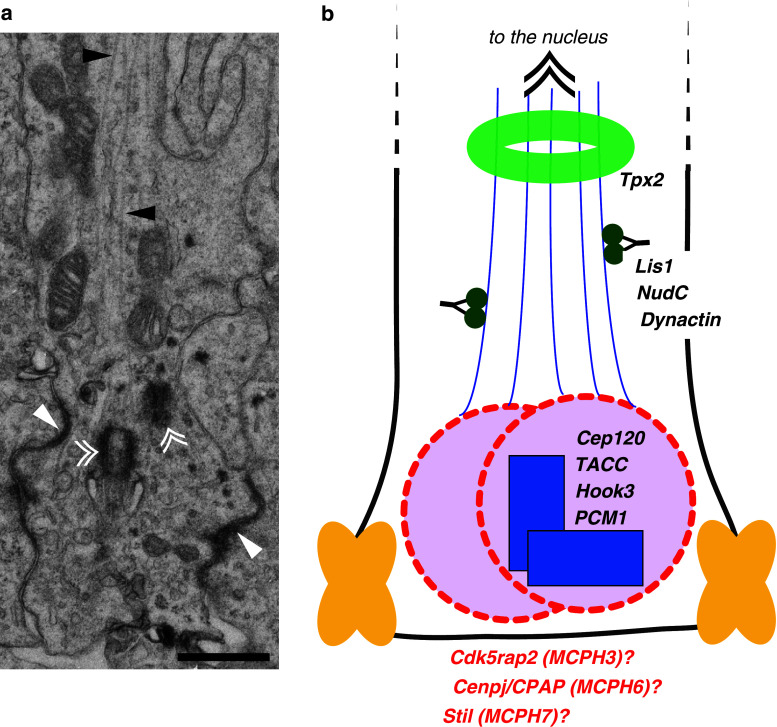
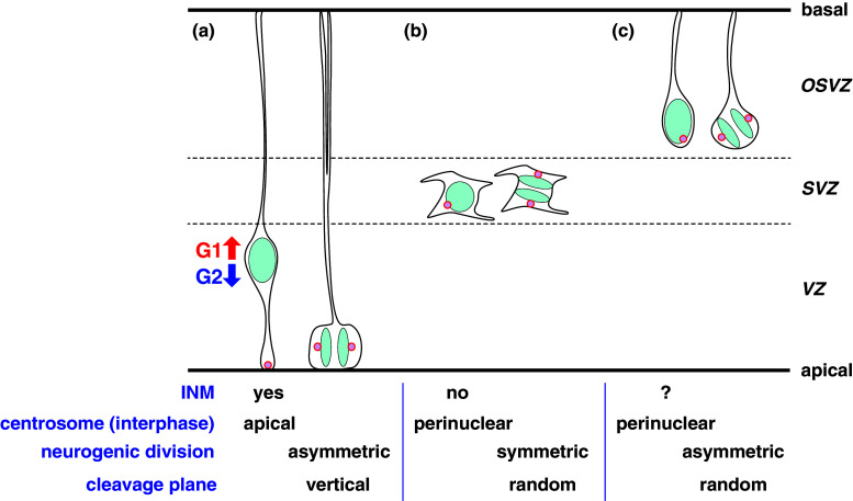
Interkinetic nuclear migration (INM) is an oscillatory nuclear movement that is synchronized with the progression of the cell cycle. The efforts of several researchers, following the first report of INM in 1935, have revealed many of the molecular mechanisms of this fascinating phenomenon linking the timing of the cell cycle and nuclear positioning in tissue. Researchers are now faced with a more fundamental question: is INM important for tissue, particularly brain, development? In this review, I summarize the current understanding of the regulatory mechanisms governing INM, investigations involving several different tissues and species, and possible explanations for how nuclear movement affects cell-fate determination and tissue formation.


Figures

Fig. 1
Fig. 1
A scheme of INM and proposed driving forces involved in INM. a The direction of nuclear movement in INM. In the G1 phase, the nucleus translocates from the apical to the basal surface (red arrow), whereas in the G2 phase, the direction is reversed (blue arrow). The small red arrow indicates the stochastic, ratcheting movement of the nucleus (see text). b Reported models for nuclear translocation during INM. Primary driving force for the nuclear migration in the G1 phase (red arrow) or G2 phase (blue arrow) is indicated close to each arrow: (i) Kif3/Dynein [18], (ii) Myosin II/Dynein [19], (iii) stochastic/Myosin II [21, 22] and (iv) displacement/Dynein [20]. For details of each model, see the text. Light blue ellipse nucleus, Thin lines attached to nucleus microtubules
Fig. 2
Fig. 2
The centrosome–microtubule complex in apical progenitor cells. a Electron micrograph of an apical endfoot in an apical progenitor cell in the mouse cortex (E14.5). The white arrowhead marks the apical junction. Double arrowheads mark the centriole. The black arrowhead marks the microtubules. Note that the microtubules run along the apical-basal axis. For specimen-preparation details, see [20]. Bar 0.5 µm. b Scheme of the centrosomemicrotubule complex. Proteins known to affect basal-to-apical nuclear migration and MCPH gene products that localize to the centrosome during interphase are shown. Yellow indicates the junctional complexes. Blue indicates the centrioles. Pink indicates the centrosome. Deep green indicates dynein. Blue line marks the microtubules. Light green marks Tpx2 accumulation on the microtubules
Fig. 3
Fig. 3
A scheme of neural progenitor cell subtypes. a Neuroepithelial/radial glial cell (apical progenitor), b basal progenitor (intermediate progenitor, non-surface dividing cell), c OSVZ progenitor (outer radial glial cell). In each cell type, interphase (left) and M phase (right) are shown. The existence of INM, the centrosome position during interphase, the type of neurogenic division and the cleavage plane orientation to the apical surface in the M phase are shown. Blue indicates the nucleus. Pink indicates the centrosomes. VZ ventricular zone, SVZ subventricular zone, OSVZ outer subventricular zone

References

    1. Sauer FC. Mitosis in the neural tube. J Comp Neurol. 1935;62:377–405. doi: 10.1002/cne.900620207. - DOI
    1. Sauer ME, Chittenden AC. Deoxyribonucleic acid content of cell nuclei in the neural tube of the chick embryo: evidence for intermitotic migration of nuclei. Exp Cell Res. 1959;16:1–6. doi: 10.1016/0014-4827(59)90189-2. - DOI - PubMed
    1. Sauer ME, Walker BE. Radioautographic study of interkinetic nuclear migration in the neural tube. Proc Soc Exp Biol Med. 1959;101:557–560. - PubMed
    1. Sidman RL, Miale IL, Feder N. Cell proliferation and migration in the primitive ependymal zone: an autoradiographic study of histogenesis in the nervous system. Exp Neurol. 1959;1:322–333. doi: 10.1016/0014-4886(59)90024-X. - DOI - PubMed
    1. Fujita S. Kinetics of cellular proliferation. Exp Cell Res. 1962;28:52–60. doi: 10.1016/0014-4827(62)90311-7. - DOI - PubMed

Publication types

LinkOut - more resources