. 2012 May;159(1):311-20.
doi: 10.1104/pp.111.191940. Epub 2012 Mar 13.

Barley ROP binding kinase1 is involved in microtubule organization and in basal penetration resistance to the barley powdery mildew fungus

Affiliations

Barley ROP binding kinase1 is involved in microtubule organization and in basal penetration resistance to the barley powdery mildew fungus

Christina Huesmann et al. Plant Physiol. 2012 May.

Abstract

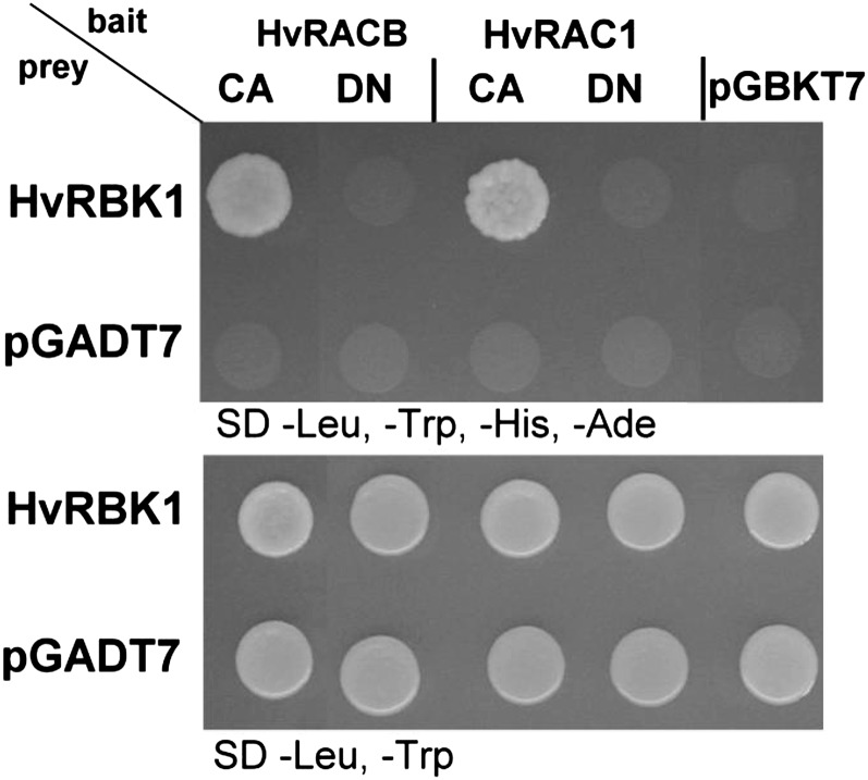
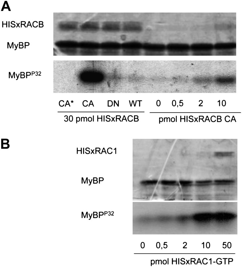
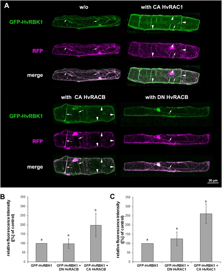
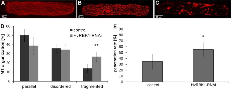
Certain plant receptor-like cytoplasmic kinases were reported to interact with small monomeric G-proteins of the RHO of plant (ROP; also called RAC) family in planta and to be activated by this interaction in vitro. We identified a barley (Hordeum vulgare) partial cDNA of a ROP binding protein kinase (HvRBK1) in yeast (Saccharomyces cerevisiae) two-hybrid screenings with barley HvROP bait proteins. Protein interaction of the constitutively activated (CA) barley HvROPs CA HvRACB and CA HvRAC1 with full-length HvRBK1 was verified in yeast and in planta. Green fluorescent protein-tagged HvRBK1 appears in the cytoplasm and nucleoplasm, but CA HvRACB or CA HvRAC1 can recruit green fluorescent protein-HvRBK1 to the cell periphery. Barley HvRBK1 is an active kinase in vitro, and activity is enhanced by CA HvRACB or GTP-loaded HvRAC1. Hence, HvRBK1 might act downstream of active HvROPs. Transient-induced gene silencing of barley HvRBK1 supported penetration by the parasitic fungus Blumeria graminis f. sp. hordei, suggesting a function of the protein in basal disease resistance. Transient knockdown of HvRBK1 also influenced the stability of cortical microtubules in barley epidermal cells. Hence, HvRBK1 might function in basal resistance to powdery mildew by influencing microtubule organization.


Figures

Figure 1.
Figure 1.
Unrooted phylogenetic tree (best scoring tree after the bootstraps) of predicted RLCKs of the Arabidopsis RLCKVIA family (At5G57670 = AtRLCK VIA1, At2G188900 = AtRLCK VIA2, At5G65530 = AtRLCK VIA3, At5G10520 = AtRLCK VIA4, At5G35960 = AtRLCK VIA5, At3G05140 = AtRLCK VIA6, At5G18910 = AtRLCK VIA7), M. truncatula MtRRKs (MtRRK1, MtRRK2), barley HvRBK1, rice OsPERK1, and the predicted transmembrane receptor-like kinase AtPERK1 (AT3G24550) taken as an outgroup. Numbers indicate support values after bootstraps.
Figure 2.
Figure 2.
Targeted YTH assay of HvRBK1 and barley HvROPs in yeast. Targeted YTH assay in yeast strain AH109 cotransformed with HvRBK1 in the pGADT7 prey vector and barley HvROPs in pGBKT7 bait vector. Bait and prey interaction allows growth on selective synthetic dextrose media without −Leu, −Trp, −His, and adenine (−Ade). No growth was observed on selective media when yeast was cotransformed with expression constructs of HvROPs and the empty vector pGADT7 or with HvRBK1 and empty vector pGBKT7. As control for successful cotransformation, yeast cells were dropped on selective synthetic dextrose media −Leu, −Trp (bottom picture).
Figure 3.
Figure 3.
Active HvRACB/HvRAC1 GTPases increase the activity of HvRBK1 in vitro. The in vitro myelin basic protein (MyBP) phosphorylating activity of HvRBK1 is shown in the presence of purified HIS-tagged barley HvRACB (A) and HvRAC1 (B) GTP-binding proteins. A, HISxHvRACB was added to the kinase reaction in CA, DN, and wild-type (WT) forms (30 pmol each). As a negative control (CA*), the same amount of the CA HvRACB GTPase was also added to a reaction mixture not containing the kinase protein (CA*). Moreover, kinase reactions were carried out in the presence of various CA HvRACB GTPase amounts (0–10 pmol). Protein loading is shown by Coomassie Brilliant Blue staining of the proteins in the kinase reaction after their separation in a polyacrylamide gel (top image), whereas kinase activity is demonstrated by the autoradiographic detection of radioactive MyBPP32 in the same gel (bottom image). B, HISxHvRAC1 was added to the kinase reaction at the indicated concentrations after loading the wild-type protein with GTP. The top image shows the result of the CBB staining and the bottom one the autoradiography of the gel used for the separation of proteins in the kinase reactions.
Figure 4.
Figure 4.
Recruitment of GFP-HvRBK1 by HvROPs in barley epidermal cells. A, Subcellular localization of GFP-HvRBK1 in epidermal cells of barley. A, Confocal laser-scanning micrographs of barley epidermal cell expressing GFP-HvRBK1 (green) and RFP (magenta). Soluble RFP was cotransformed as marker for cytoplasmic and nuclear localization. White color in the merged channels demonstrates similar localization of GFP-HvRBK1 and RFP in cytoplasmic strands (arrows). GFP-HvRBK1 was expressed alone (−/−) or together with the unlabeled HvROPs CA HvRACB, CA HvRAC1, or DN HvRACB. Coexpression of GFP-HvRBK1 and CA HvRACB or CA HvRAC1 results in recruitment of GFP-HvRBK1 to the cell periphery/plasma membrane (arrowheads) as demonstrated by color separation and the green cell periphery in the fluorescent merged pictures. DN HvRACB does not alter cytoplasmic localization of GFP-HvRBK1. Pictures are maximum projections of 20 to 30 optical sections at 2-μm increments. B and C, Fluorescence intensity of GFP-HvRBK1 at the cell periphery is greatly modified upon coexpression of different HvROPs. To quantify recruitment of GFP-HvRBK1 by CA HvRACB and CA HvRAC1, both proteins were coexpressed with GFP-HvRBK1 and mCherry as a marker for cytoplasmic and nuclear localization. As negative control, CA HvROP variants were replaced by DN HvRACB or DN HvRAC1. Additionally, the GFP-HvRBK1 was expressed with mCherry and empty vector as control. Mean pixel intensity was measured at the cell periphery and normalized against mCherry pixel intensity in the nucleus. Columns show means of three independent experiments with 95% confidence intervals as error bars. Intensities were significantly different between the CA HvRACB or CA HvRAC1 expressing cells versus the respective controls after ANOVA (Tukey test, P < 0.05, as indicated by different letters in the figure).
Figure 5.
Figure 5.
Influences of TIGS of HvRBK1 on fungal penetration success and MT organization in epidermal cells of barley. Epidermal cells were transiently transformed with the MT marker RFP-HvMAGAP1 to image typical MT arrays in epidermal cells. A, Parallel arrangement of MTs. B, Disordered/randomized arrangement of MTs. C, Fragmented MTs. D, Frequencies of cells falling into the categories of different MT arrays (A–C). Columns represent means of four independent experiments (50 cells for each plasmid combination were investigated per experiment), with significantly more fragmented MTs after TIGS of HvRBK1. We counted MT arrays after projections of 20 to 25 optical sections at 2-μm increments with the MT marker RFP-HvMAGAP1-Cter. Similar results were obtained with two alternative MT markers (Supplemental Fig. S5). E, TIGS of HvRBK1, when compared with empty TIGS vector control, led to enhanced fungal penetration rate as indicated by more haustoria-containing cells per total cells attacked by B. graminis f. sp. hordei. Columns show the mean of five independent biological experiments. Error bars show sd of the mean (two-sided Student’s t test; *, P < 0.05 and **, P < 0.01). [See online article for color version of this figure.]

Similar articles

Cited by

References

    1. Baluska F, Bacigálová K, Oud JL, Hauskrecht M, Kubica G. (1995) Rapid reorganization of microtubular cytoskeleton accompanies early changes in nuclear ploidy and chromatin structure in postmitotic cells of barley leaves infected with powdery mildew. Protoplasma 185: 140–151
    1. Berken A, Wittinghofer A. (2008) Structure and function of Rho-type molecular switches in plants. Plant Physiol Biochem 46: 380–393 - PubMed
    1. Bustelo XR, Sauzeau V, Berenjeno IM. (2007) GTP-binding proteins of the Rho/Rac family: regulation, effectors and functions in vivo. Bioessays 29: 356–370 - PMC - PubMed
    1. Chen L, Hamada S, Fujiwara M, Zhu T, Thao NP, Wong HL, Krishna P, Ueda T, Kaku H, Shibuya N, et al. (2010a) The Hop/Sti1-Hsp90 chaperone complex facilitates the maturation and transport of a PAMP receptor in rice innate immunity. Cell Host Microbe 7: 185–196 - PubMed
    1. Chen L, Shiotani K, Togashi T, Miki D, Aoyama M, Wong HL, Kawasaki T, Shimamoto K. (2010b) Analysis of the Rac/Rop small GTPase family in rice: expression, subcellular localization and role in disease resistance. Plant Cell Physiol 51: 585–595 - PubMed

Publication types

MeSH terms

LinkOut - more resources