. 2012 Mar 13:3:735.
doi: 10.1038/ncomms1734.

Activation of canonical Wnt signalling is required for TGF-β-mediated fibrosis

Affiliations
Free PMC article

Activation of canonical Wnt signalling is required for TGF-β-mediated fibrosis

Alfiya Akhmetshina et al. Nat Commun. .
Free PMC article

Abstract

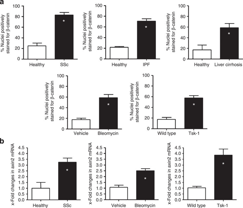
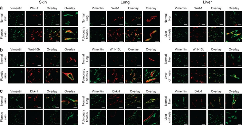
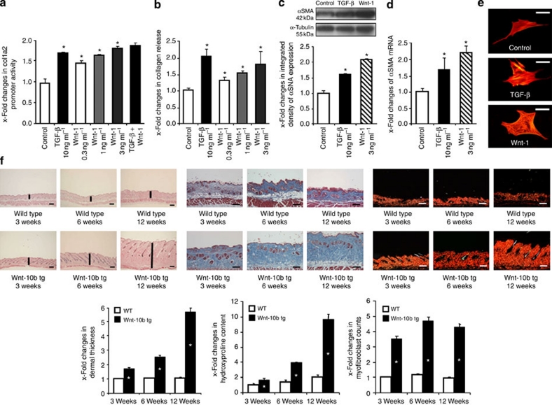
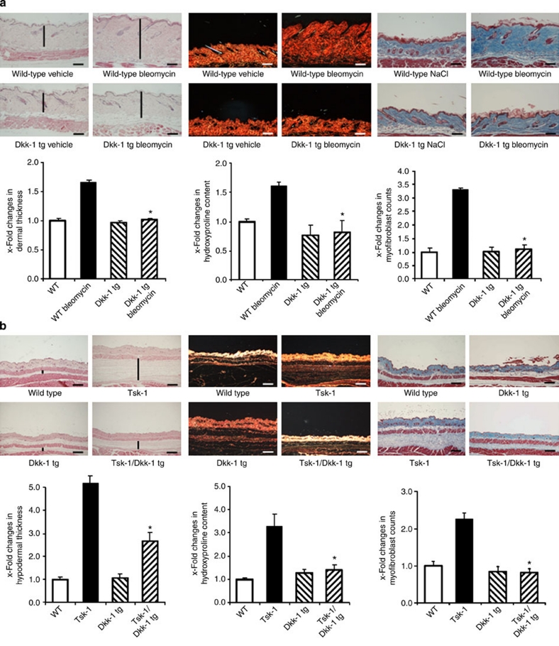
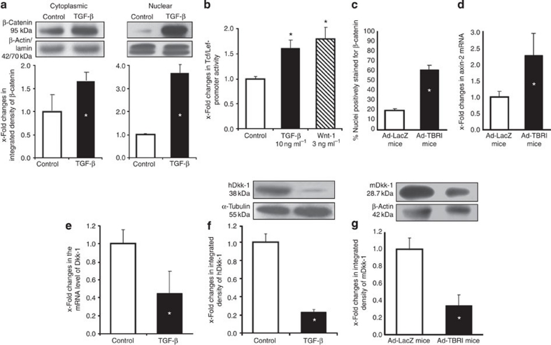
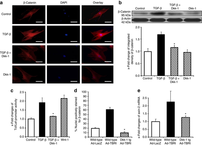
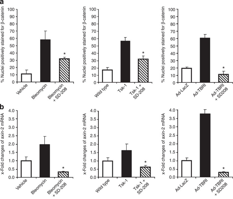
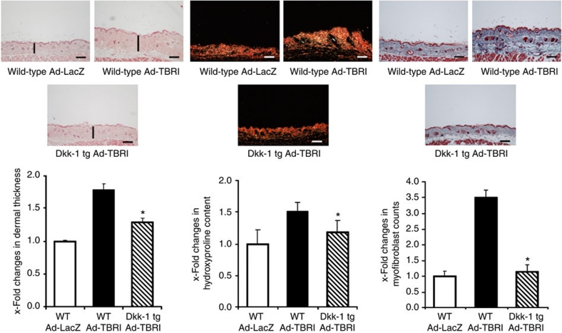
The transforming growth factor-β (TGF-β) signalling pathway is a key mediator of fibroblast activation that drives the aberrant synthesis of extracellular matrix in fibrotic diseases. Here we demonstrate a novel link between transforming growth factor-β and the canonical Wnt pathway. TGF-β stimulates canonical Wnt signalling in a p38-dependent manner by decreasing the expression of the Wnt antagonist Dickkopf-1. Tissue samples from human fibrotic diseases show enhanced expression of Wnt proteins and decreased expression of Dickkopf-1. Activation of the canonical Wnt pathway stimulates fibroblasts in vitro and induces fibrosis in vivo. Transgenic overexpression of Dickkopf-1 ameliorates skin fibrosis induced by constitutively active TGF-β receptor type I signalling and also prevents fibrosis in other TGF-β-dependent animal models. These findings demonstrate that canonical Wnt signalling is necessary for TGF-β-mediated fibrosis and highlight a key role for the interaction of both pathways in the pathogenesis of fibrotic diseases.


Figures

Figure 1
Figure 1. Canonical Wnt signalling is activated in fibrotic diseases.
(a) β-catenin, the central signalling component of the canonical Wnt pathway, accumulated in the nuclei of fibroblasts in samples from patients with SSc (n=12 for SSc and n=10 for normal skin), IPF (n=8 for IPF and n=6 for non-fibrotic lung) and liver cirrhosis (n=6 for liver cirrhosis and n=6 for non-fibrotic liver). Nuclear accumulation of β-catenin in dermal fibroblasts was also observed in the experimental models of bleomycin-induced dermal fibrosis and in Tsk-1 mice (n=10 each). A minimum of 100 nuclei per sample was counted. (b) The mRNA levels of the Wnt target gene axin-2 were significantly increased in systemic sclerosis patients, bleomycin challenged mice and Tsk-1 mice (n=8 each). * Indicates P-values of less than 0.05 (analysed using the Mann–Whitney U-test) as compared with healthy volunteers or with non-fibrotic control mice, respectively. All data are expressed as mean ± s.e.m.
Figure 2
Figure 2. Wnt-1 and Wnt-10b are upregulated in fibrotic diseases and Dkk-1 is downregulated.
(a) Confocal microscopy with double staining for Wnt-1 and vimentin demonstrated an overexpression of Wnt-1, in particular, in vimentin-positive cells in fibrotic skin of patients with SSc, in IPF and in liver cirrhosis. (b) Wnt-10b was also overexpressed in SSc, IPF and liver cirrhosis. (c) In parallel to the upregulation of Wnt-1 and Wnt-10b, a prominent decrease of the endogenous antagonist Dkk-1 was observed in fibroblasts in fibrotic diseases. Representative examples from 12 patients with SSc, 10 samples of non-fibrotic skin, 10 patients with IPF, 6 control lungs, 8 patients with liver cirrhosis and 8 control livers are shown. Scale bars, 20 or 5 μm for the outer right image of each panel, respectively.
Figure 3
Figure 3. Activation of the canonical Wnt pathway induces fibrosis.
(a) Recombinant Wnt-1 stimulated the activity of col1a2 promoter constructs (n=6). (b) Incubation with recombinant Wnt-1 increased the release of collagen protein by cultured fibroblasts in a dose-dependent manner (n=6). (c) Wnt-1 induced the expression of αSMA protein (n=5). (d) Incubation with Wnt-1 increased the mRNA levels of αSMA (n=5). (e) Wnt-1 stimulated the formation of stress fibres in cultured fibroblasts (n=6). Of note, the stimulatory effects of Wnt-1 on fibroblasts were comparable with those of TGF-β. (f) Transgenic overexpression of Wnt-10b induced dermal fibrosis in mice. The dermal thickness, the hydroxyproline content and the numbers of myofibroblasts were already increased in Wnt-10b tg mice at an age of 3 weeks and increased further with age (n ≥ 5 for all groups). Representative haematoxylin and eosin-, trichrome- and Sirius Red-stained sections of wild-type (WT) and Wnt-10b transgenic (Wnt-10b tg) mice at ages of 3, 6 and 12 weeks are shown (horizontal scale bars, 100 μm). Black vertical bars indicate the dermal thickness. * Indicates P-values of less than 0.05 as compared with mock-treated fibroblasts (ae, analysed with the Wilcoxon signed-rank test) or compared with wild-type littermates (f, analysed using the Mann–Whitney U-test), respectively. All data are expressed as mean ± s.e.m.
Figure 4
Figure 4. Inhibition of canonical Wnt signalling by Dkk-1 prevents experimental fibrosis.
(a) Dkk-1 transgenic (Dkk-1 tg) mice were resistant to bleomycin-induced fibrosis. Haematoxylin and eosin-, Sirius Red- and trichrome-stained sections are shown (horizontal scale bars, 100 μm). Challenge with bleomycin induced dermal fibrosis with increased dermal thickening, accumulation of hydroxyproline and myofibroblast differentiation in wild-type mice, but not in Dkk-1 tg littermates. In contrast to wild-type mice, these outcomes did not differ between Dkk-1 tg mice challenged with bleomycin and Dkk-1 tg mice injected with NaCl (n ≥ 6 for all groups). (b) Dkk-1 prevented fibrosis in Tsk-1 mice. The histological changes induced by the Tsk-1 mutation in the fibrillin-1 gene are ameliorated by transgenic overexpression of Dkk-1, as shown in representative haematoxylin and eosin-, Sirius Red- and trichrome-stained sections (horizontal scale bars, 200 μm). Hypodermal thickening, hydroxyproline content and myofibroblast counts were all significantly reduced in Tsk-1/Dkk-1, as compared with Tsk-1 littermates not carrying the Dkk-1 transgene (n ≥ 5 for all groups). Vertical black bars in these images indicate the dermal or hypodermal thickness. *Indicates P-values of less than 0.05 as compared with wild-type mice challenged with bleomycin (a) and with Tsk-1 mice not transgenic for Dkk-1 (b), respectively, as analysed using the Mann–Whitney U-test. All data are expressed as mean ± s.e.m.
Figure 5
Figure 5. TGF-β activates canonical Wnt signalling by decreasing Dkk-1.
(a) TGF-β induces nuclear accumulation of β-catenin in cultured human fibroblasts as shown by Western blot from cytoplasmic and nuclear extracts (n=4). (b) TGF-β stimulates the activity of a TCF/Lef reporter construct (n=8). (c) Adenoviral overexpression of TBRIact in the skin of mice (Ad-TBRI) induced nuclear accumulation of β-catenin in fibroblasts as compared with control mice infected with adenovirus encoding for LacZ (Ad-LacZ) (n ≥ 6). (d) Ad-TBRI uprgeulated the mRNA levels of the Wnt target gene axin-2 (n ≥ 6). (e) Incubation with TGF-β reduced the mRNA levels of Dkk-1 in cultured fibroblasts (n=7). (f) TGF-β decreased the protein levels of Dkk-1 in cultured fibroblasts (n=5). (g) Decreased levels of Dkk-1 were also observed in the skin of Ad-TBRI mice by western blot (n=4). Representative bands and the mean results of the densiometric quantification are shown in a, f and g. * Indicates P-values of less than 0.05 as compared with unstimulated cells (a, b, e and f, as analysed with the Wilcoxon signed-rank test) and with Ad-LacZ mice (c, d and g, as analysed using the Mann–Whitney U-test). All data are expressed as mean ± s.e.m.
Figure 6
Figure 6. Dkk-1 prevented the stimulatory effects of TGF-β on canonical Wnt signalling.
(a) Recombinant Dkk-1 abrogated the TGF-β induced nuclear accumulation of β-catenin as assessed by immunofluorescence (n=6). (b) The inhibitory effects of Dkk-1 on the TGF-β-induced nuclear accumulation of β-catenin were confirmed by western blot (n=4). (c) Dkk-1 prevented the TGF-β-induced activation of a TCF/Lef reporter construct in vitro. Recombinant Wnt-1 was used as a positive control (n=8). (d) Nuclear accumulation of β-catenin was observed in wild-type mice infected with adenovirus encoding for TBRIact (Ad-TBR), but not in Dkk-1 transgenic mice (Dkk-1 tg; n ≥ 5 for all groups). (e) Ad-TBRI increased the mRNA levels of axin-2 in wild-type mice, but not Dkk-1 tg mice (n ≥ 5 for all groups). * Indicates P-values of less than 0.05 as compared with cells stimulated with TGF-β (b,c) or with wild-type mice infected with Ad-TBRI (d,e). All results were analysed using the Wilcoxon signed-rank test. All data are expressed as mean ± s.e.m.
Figure 7
Figure 7. Inhibition of TGF-β signalling reduces the activation of canonical Wnt in experimental fibrosis.
(a) Treatment with SD-208, a selective inhibitor of TBRI, significantly decreased the nuclear accumulation of β-catenin in fibroblasts in bleomycin-induced dermal fibrosis, in Tsk-1 mice and in TBRIact induced fibrosis (n ≥ 5 for all groups in all models). (b) SD-208 completely prevented the increase in the mRNA levels of axin-2 in these mouse models (n ≥ 5 for all groups in all models). * Indicates P-values of less than 0.05 (as analysed with the Mann–Whitney U-test) compared with bleomycin treated-, Tsk-1, or TBRIact mice without SD-208. All data are expressed as mean ± s.e.m.
Figure 8
Figure 8. Inhibition of canonical Wnt signalling by Dkk-1 prevents Ad-TBRIact-induced fibrosis.
Intradermal infection with adenoviruses encoding for TBRIact (Ad-TBRI) induced dermal fibrosis in wild-type mice, but not in Dkk-1 transgenic (Dkk-1 tg) littermates. Representative histological sections stained with haematoxylin and eosin, Sirius Red and trichrome are shown (horizontal scale bar, 100 μm). The dermal thickening observed in wild-type mice on infection with Ad-TBRI was significantly ameliorated in Dkk-1 transgenic (Dkk-1 tg) mice. The Ad-TBRI-mediated increases in hydroxyproline content and in the number of myofibroblasts were also reduced in Dkk-1 tg mice (n ≥ 5 for all groups). * Indicates P-values of less than 0.05 (as analysed using the Mann–Whitney U-test) compared with wild-type mice infected with Ad-TBRI. All data are expressed as mean ± s.e.m.

References

    1. Wynn T. A. Cellular and molecular mechanisms of fibrosis. J. Pathol. 214, 199–210 (2008). - PMC - PubMed
    1. Gabrielli A., Avvedimento E. V. & Krieg T. Scleroderma. N. Engl. J. Med. 360, 1989–2003 (2009). - PubMed
    1. Charles C., Clements P. & Furst D. E. Systemic sclerosis: hypothesis-driven treatment strategies. Lancet 367, 1683–1691 (2006). - PubMed
    1. Strieter R. M. & Mehrad B. New mechanisms of pulmonary fibrosis. Chest 136, 1364–1370 (2009). - PMC - PubMed
    1. Verrecchia F. & Mauviel A. Transforming growth factor-beta and fibrosis. World J. Gastroenterol. 13, 3056–3062 (2007). - PMC - PubMed

Publication types

MeSH terms

Substances