Comparative Study
. 2012 Mar 13:3:741.
doi: 10.1038/ncomms1730.

Fast and ultrasensitive method for quantitating prion infectivity titre

Affiliations
Comparative Study

Fast and ultrasensitive method for quantitating prion infectivity titre

Natallia Makarava et al. Nat Commun. .

Abstract

Bioassay by end-point dilution has been used for decades for routine determination of prion infectivity titre. Here we show that the new protein misfolding cyclic amplification with beads (PMCAb) technique can be used to estimate titres of the infection-specific forms of the prion protein with a higher level of precision and in 3-6 days as opposed to 2 years, when compared with the bioassay. For two hamster strains, 263 K and SSLOW, the median reactive doses determined by PCMAb (PMCAb(50)) were found to be 10(12.8) and 10(12.2) per gram of brain tissue, which are 160- and 4,000-fold higher than the corresponding median infectious dose (ID(50)) values measured by bioassay. The 10(2)- to 10(3)-fold differences between ID(50) and PMCAb(50) values could be due to a large excess of PMCAb-reactive prion protein seeds with little or no infectivity. Alternatively, the differences between ID(50) and PMCAb(50) could be due to higher rate of clearance of infection-specific prion protein seeds in animals versus PMCAb reactions. A well-calibrated PMCAb reaction can be an efficient and cost-effective method for the estimation of infection-specific prion protein titre.


Figures
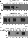
Figure 1
Figure 1. End-point sPMCAb titration
10% scrapie brain material from SSLOW- (a) or 263K infected animals (b, c) were diluted in ten fold serial steps, then aliquots from each dilution were used to seed serial PMCAb reactions. Example of a Western blot of sPMCA products amplified for 6 rounds is shown in plots a and b. Panel c shows products of PMCAb reactions seeded with four dilutions and amplified for 2, 3 or 6 serial rounds. The dilutions shown for PMCAb reactions were normalized per gram of brain tissue, to correct for differences in assay volumes used for PMCAb (100 μl) and bioassay (50 μl).
Figure 2
Figure 2. Testing the specificity of PrPSc detection in sPMCAb
sPMCAb reactions were seeded with 10% NBH from 700 day old S. hamsters (lanes 5 and 6), recombinant PrP (rPrP) amyloid fibrils type I not subjected to annealing (lane 7); fibrils type I subjected to annealing in Triton (lane 8), BSA (lane 9) or NBH (lanes 10, 11 and 12); with rPrP amyloid fibrils type II (lane 13); or 1011-fold diluted SSLOW brain material (lane14). Four non-seeded reactions are shown in lanes 1, 2, 3 and 4. Six rounds of sPMCAb were conducted for each seeded condition and non-seeded reaction. The final concentration of rPrP amyloid fibrils in sPMCAb reactions was 10 μg/ml. rPrP fibrils were prepared using Syrian Hamster full-length rPrP; fibrils type I were prepared in 1M GdnHCl and 3M urea as described by Bocharova et al ; fibrils type II were prepared in 0.5 M GdnHCl as described by Sun et al . rPrP fibrils type I were subjected to annealing in 1% Tritorn X-100, 5 mg/ml BSA, or 5% NBH as described by Bocharova et al. and Makarava et al. . After six sPMCAb rounds, all samples were treated with PK as described in Methods; Western blots were stained with 3F4. To rigorously test the specificity of sPMCAb for detecting PrPSc, multiple experiments were performed using four Misonix S-4000 sonicators over a two year period. Several independently prepared stocks of rPrP amyloid fibrils of each type were produced using rPrP from several independent purifications. All results on sPMCAb seeding by rPrP fibrils were negative, confirming that sPMCAb is highly specific for PrPSc.
Figure 3
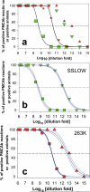
Figure 3. Correlation of prion infectivity titer by end-point titration bioassay with PMCAb activity
Brain homogenate materials from Syrian Hamsters infected with SSLOW (green symbols) or 263K (red symbols) were subjected to 10-fold serial dilution, then each dilution was analyzed by animal bioassay or serial PMCAb. Percentage of animals infected (squares) or giving positive PMCAb reactions (triangles) is plotted as a function of dilution. (a) Analysis of the data using Poisson function, where the solid curves represent the results of nonlinear least-square best fit. Analysis of the data for SSLOW (b) or 263K (c) using sigmoidal function. The solid curves represent the results of nonlinear least-square best fit and the blue curves represent 95% confidence intervals. ID50 and PMCAb50 values were calculated from the results of fitting.

References

    1. Wadsworth JD, et al. Tissues distribution of protease resistant prion protein in variant Creutzfeldt-Jakob disease using a highly sensitive immunobloting assay. Lancet. 2001;358:171–180. - PubMed
    1. Safar JG, et al. Measuring prions causing bovine spongiform encephalopathy or chronic wasting disease by immunoassays and transgenic mice. Nat. Biotechnol. 2002;20:1147–1150. - PubMed
    1. Edgeworth JA, et al. Detection of prion infection in variant Creutzfeldt-Jakob disease: a blood-based assay. Lancet. 2011;377:487–493. - PubMed
    1. Gregori L, et al. A sensitive and quantitative assay for normal PrP in plasma. J. Virol Methods. 2008;149:251–259. - PubMed
    1. Silveira JR, et al. The most infectious prion protein particles. Nature. 2005;437:257–261. - PMC - PubMed

Publication types