. 2012 May;153(5):2311-22.
doi: 10.1210/en.2011-1452. Epub 2012 Mar 13.

Growth hormone-induced JAK2 signaling and GH receptor down-regulation: role of GH receptor intracellular domain tyrosine residues

Affiliations

Growth hormone-induced JAK2 signaling and GH receptor down-regulation: role of GH receptor intracellular domain tyrosine residues

Luqin Deng et al. Endocrinology. 2012 May.

Abstract

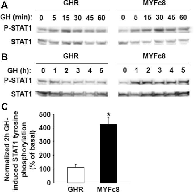
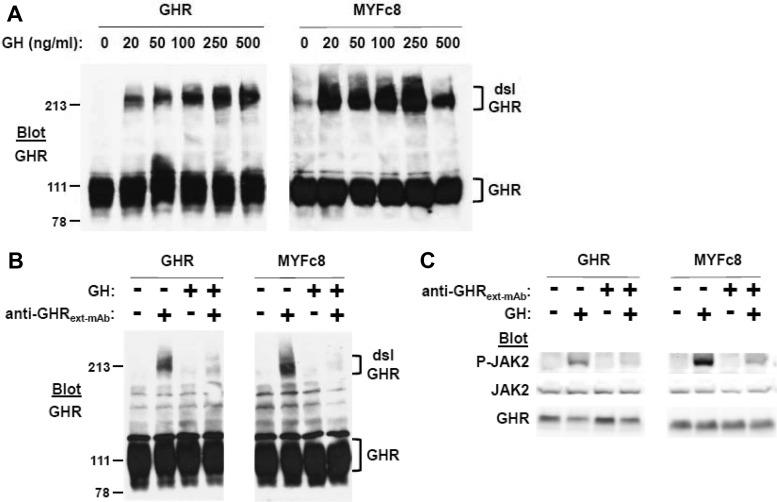
GH receptor (GHR) mediates important somatogenic and metabolic effects of GH. A thorough understanding of GH action requires intimate knowledge of GHR activation mechanisms, as well as determinants of GH-induced receptor down-regulation. We previously demonstrated that a GHR mutant in which all intracellular tyrosine residues were changed to phenylalanine was defective in its ability to activate signal transducer and activator of transcription (STAT)5 and deficient in GH-induced down-regulation, but able to allow GH-induced Janus family of tyrosine kinase 2 (JAK2) activation. We now further characterize the signaling and trafficking characteristics of this receptor mutant. We find that the mutant receptor's extracellular domain conformation and its interaction with GH are indistinguishable from the wild-type receptor. Yet the mutant differs greatly from the wild-type in that GH-induced JAK2 activation is augmented and far more persistent in cells bearing the mutant receptor. Notably, unlike STAT5 tyrosine phosphorylation, GH-induced STAT1 tyrosine phosphorylation is retained and augmented in mutant GHR-expressing cells. The defective receptor down-regulation and persistent JAK2 activation of the mutant receptor do not depend on the sustained presence of GH or on the cell's ability to carry out new protein synthesis. Mutant receptors that exhibit resistance to GH-induced down-regulation are enriched in the disulfide-linked form of the receptor, which reflects the receptor's activated conformation. Furthermore, acute GH-induced internalization, a proximal step in down-regulation, is markedly impaired in the mutant receptor compared to the wild-type receptor. These findings are discussed in the context of determinants and mechanisms of regulation of GHR down-regulation.


Figures

Fig. 1.
Fig. 1.
Altered kinetics of GH signaling in MYFc8 cells vs. GHR cells. A and B, Acute GH-induced JAK2 and STAT5 signaling. A, Serum-starved GHR and MYFc8 cells were treated with vehicle or GH (500 ng/ml; 15 min). Detergent extracts were resolved by SDS-PAGE and sequentially immunoblotted to detect phosphorylated and total JAK2 and phosphorylated and total STAT5. The figure shown is representative of six such experiments. B, Experiments (n = 6) such as those shown in panel A, but with 250 ng/ml GH, were analyzed denstiometrically. In each case, P-JAK2 signal was normalized for JAK2 signal and expressed as the normalized GH-induced JAK2 activation in MYFc8 cells relative to that in GHR cells. *, P < 0.05 between conditions. C and D, GH signaling time course. C, Serum-starved GHR and MYFc8 cells were treated with vehicle for 60 min (left panel) or 5 h (right panel) or with GH (250 ng/ml) for the indicated durations before detergent extraction, SDS-PAGE, and sequential immunoblotting, as indicated. Note that blots in the left and right panels were actually contiguous (run on the same gel), but are separated for ease of visualization; exposures are thus processed identically and are comparable in the left and right panels. The figure shown is representative of three such experiments. D, Experiments (n = 4) such as those shown in panel C were performed for MYFc8 cells and analyzed densitometrically as in panel B. Normalized GH-induced JAK2 activation is expressed at each GH time point relative to the normalized signal obtained after 1 h of GH exposure. E, GH pulse and washout. Serum-starved GHR and MYFc8 cells were treated with vehicle (−) or GH (250 ng/ml) for 15 min, after which the GH was washed out and cells were incubated thereafter for 0, 15, 30, or 60 min, as indicated. Cells were then detergent extracted, and lysates were resolved by SDS-PAGE, followed by sequential immunoblotting, as indicated. Note that blots in the left and right panels were actually contiguous (run on the same gel), but are separated for ease of visualization; exposures are thus processed identically and are comparable in the left and right panels. The figure shown is representative of three such experiments.
Fig. 2.
Fig. 2.
GH-induced STAT1 tyrosine phosphorylation in MYFc8 cells vs. GHR cells. A and B, Serum-starved GHR and MYFc8 cells were treated with vehicle for 60 min (A) or 5 h (B) or with GH (250 ng/ml) for the indicated durations before detergent extraction, SDS-PAGE, and sequential immunoblotting, as indicated. Note that blots in the left and right panels were actually contiguous (run on the same gel), but are separated for ease of visualization; exposures are thus processed identically and are comparable in the left and right panels. The figure shown is representative of three such experiments. C, Experiments (n = 3) such as that shown in panel B, were analyzed denstiometrically. In each case, GH treatment for 2 h was compared with control treatment. P-JAK2 signal was normalized for JAK2 signal and expressed as the normalized GH-induced JAK2 activation in MYFc8 cells relative to that in GHR cells. *, P < 0.05 between conditions.
Fig. 3.
Fig. 3.
GH-induced GHR disulfide linkage and anti-GHRext-mAb inhibition in MYFc8 cells vs. GHR cells. A, Acute GH-induced GHR disulfide linkage. Serum-starved GHR and MYFc8 cells were treated with GH (0–500 ng/ml, as indicated) for 15 min, before detergent extraction, SDS-PAGE under nonreducing conditions, and anti-GHR immunoblotting. Positions of the disulfide-linked (dsl) and non-disulfide-linked GHR are indicated by brackets. Note that blots in the left and right panels were actually contiguous (run on the same gel), but are separated for ease of visualization; exposures are thus processed identically and are comparable in the left and right panels. The figure shown is representative of three such experiments. B, Anti-GHRext-mAb inhibition of GH-induced GHR disulfide linkage. Serum-starved GHR and MYFc8 cells were pretreated with vehicle or anti-GHRext-mAb (18 μg/ml) for 10 min before treatment with vehicle or GH (500 ng/ml) for 15 min. Cells were then detergent extracted and lysates were resolved by SDS-PAGE under nonreducing conditions, followed by anti-GHR immunoblotting. Positions of the disulfide-linked (dsl) and non-disulfide-linked GHR are indicated by brackets. Note that blots in the left and right panels were actually contiguous (run on the same gel), but are separated for ease of visualization; exposures are thus processed identically and are comparable in the left and right panels. The figure shown is representative of three such experiments. C, Anti-GHRext-mAb inhibition of GH signaling. Serum-starved GHR and MYFc8 cells were pretreated with vehicle or anti-GHRext-mAb (18 μg/ml) for 10 min before treatment with vehicle or GH (500 ng/ml) for 15 min. Cells were then detergent extracted, and lysates were resolved by SDS-PAGE, followed by sequential immunoblotting, as indicated. Note that blots in the left and right panels were actually contiguous (run on the same gel), but are separated for ease of visualization; exposures are thus processed identically and are comparable in the left and right panels. The figure shown is representative of three such experiments.
Fig. 4.
Fig. 4.
PMA-induced GHR proteolysis in MYFc8 cells vs. GHR cells. Serum-starved GHR and MYFc8 cells were treated with PMA (0.1 μg/ml) for 0–30 min. Cells were then detergent extracted, and lysates were resolved by SDS-PAGE, followed by anti-GHR immunoblotting. The positions of GHR and the GHR remnant are indicated by brackets. Note that blots in the left and right panels were actually contiguous (run on the same gel), but are separated for ease of visualization; exposures are thus processed identically and are comparable in the left and right panels. The figure shown is representative of three such experiments.
Fig. 5.
Fig. 5.
GH-induced GHR down-regulation and effects of CHX in MYFc8 cells vs. GHR cells. A, GH-induced GHR down-regulation. Serum-starved GHR and MYFc8 cells were treated with GH (250 ng/ml) for 2 h, before detergent extraction, SDS-PAGE, and immunoblotting for GHR, as in Fig. 2C, above. GHR abundance after GH stimulation was normalized for abundance in control-treated cells for each cell type (n = 3). *, P < 0.05 between conditions. B, GH-induced GHR receptor down-regulation concentration dependance. Serum-starved GHR and MYFc8 cells were treated with GH (0–500 ng/ml, as indicated) for 1.5 h, before detergent extraction, SDS-PAGE, and sequential immunoblotting, as indicated. Note that blots in the left and right panels were actually contiguous (run on the same gel), but are separated for ease of visualization; exposures are thus processed identically and are comparable in the left and right panels. The figure shown is representative of three such experiments. C–F, Effects of CHX treatment. Serum-starved GHR and MYFc8 cells were treated with CHX (20 μg/ml) for either 60 min or 5 h, as indicated, and with GH (250 ng/ml) for the indicated durations, before detergent extraction, SDS-PAGE (under nonreducing conditions in panel F), and immunoblotting with anti-GHR (panels C and F) or sequentially as indicated (panel E). Note that blots in the left and right panels of C and F were actually contiguous (run on the same gel), but are separated for ease of visualization; exposures are thus processed identically and are comparable in the left and right panels. The figures shown are representative of three such experiments in each of panels C, E, and F. In panel D, relative abundance of GHR in GHR vs. MYFc8 cells treated with GH for 2 h compared with control treatment were densitometrically analyzed (n = 3), as in panel A above. *, P < 0.05 between conditions.
Fig. 6.
Fig. 6.
Surface biotinylation and GHR internalization assay in MYFc8 cells vs. GHR cells. A, Surface biotinylation. Serum-starved GHR and MYFc8 cells were treated with vehicle or biotin coupler, as in Materials and Methods. Cells were detergent extracted, and aliquots were precipitated with avidin beads and eluted or not precipitated before SDS-PAGE and anti-GHR immunoblotting. Note that blots in the left and right panels were actually contiguous (run on the same gel), but are separated for ease of visualization; exposures are thus processed identically and are comparable in the left and right panels. The figure shown is representative of two such experiments. B, Surface biotinylation does not affect GH-induced signaling. Serum-starved GHR cells and MYFc8 cells were treated with vehicle or biotin coupler, as in Materials and Methods, before stimulation with vehicle or GH (500 ng/ml; 15 min). Cells were detergent extracted, and extracts were resolved by SDS-PAGE and sequentially immunoblotted, as indicated. Note that blots in the left and right panels were actually contiguous (run on the same gel), but are separated for ease of visualization; exposures are thus processed identically and are comparable in the left and right panels. C, Internalization assay. Serum-starved GHR and MYFc8 cells were subjected to surface biotinylation and the internalization assay, as in Materials and Methods. GHR internalization (expressed as the % of total mature GHR detected by blotting) is plotted. (n = 4) *, P < 0.05 for GHR cells vs. MYFc8 cells at the indicated time point.

References

    1. Kaplan S. 1999. Hormonal regulation of growth and metabolic effects of growth hormone. In: Kostyo JL, Goodman HM, eds. Handbook of physiology. Chap 5 New York: Oxford University Press; 129–143
    1. Møller N, Jørgensen JO. 2009. Effects of growth hormone on glucose, lipid, and protein metabolism in human subjects. Endocr Rev 30:152–177 - PubMed
    1. Carter-Su C, Schwartz J, Smit LS. 1996. Molecular mechanism of growth hormone action. Annu Rev Physiol 58:187–207 - PubMed
    1. Frank SJ, Gilliland G, Kraft AS, Arnold CS. 1994. Interaction of the growth hormone receptor cytoplasmic domain with the JAK2 tyrosine kinase. Endocrinology 135:2228–2239 - PubMed
    1. Frank SJ, Yi W, Zhao Y, Goldsmith JF, Gilliland G, Jiang J, Sakai I, Kraft AS. 1995. Regions of the JAK2 tyrosine kinase required for coupling to the growth hormone receptor. J Biol Chem 270:14776–14785 - PubMed

Publication types

MeSH terms