. 2012 Nov;22(6):788-97.
doi: 10.1111/j.1750-3639.2012.00592.x. Epub 2012 Apr 19.

ALDH1A1 is a marker of astrocytic differentiation during brain development and correlates with better survival in glioblastoma patients

Affiliations

ALDH1A1 is a marker of astrocytic differentiation during brain development and correlates with better survival in glioblastoma patients

S Alexandra Adam et al. Brain Pathol. 2012 Nov.

Abstract

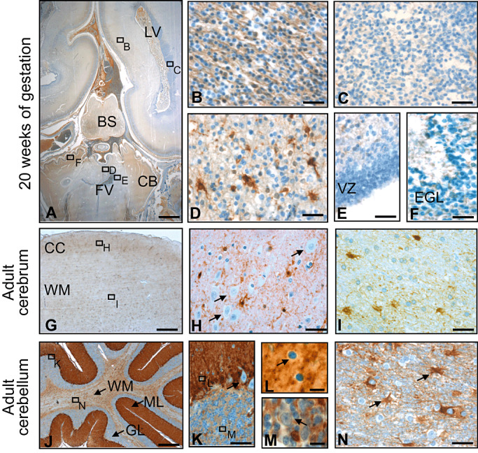
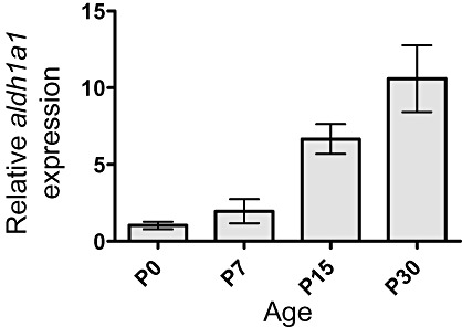
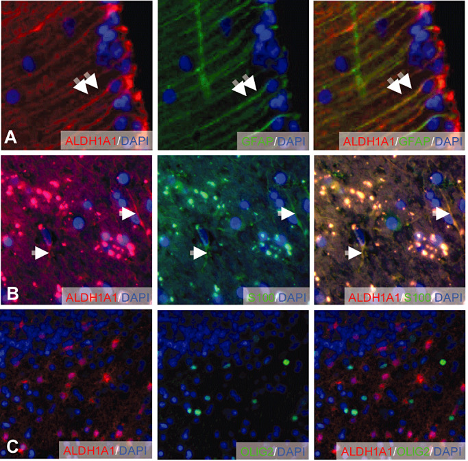
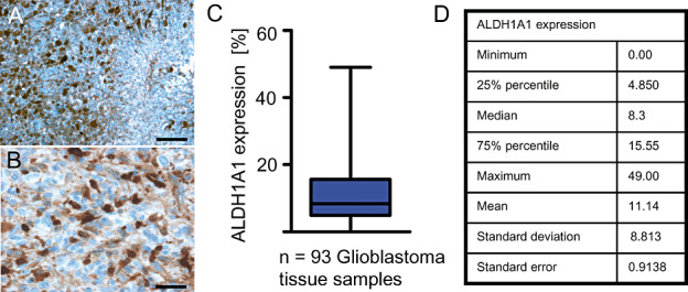
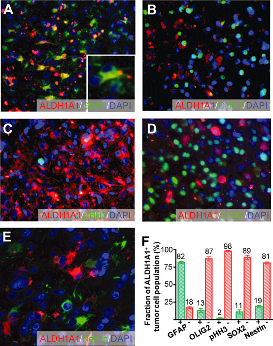
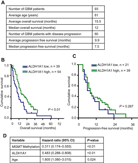
Glioblastoma is the most common malignant brain tumor and patients usually succumb to their disease within 2 years. Aldehyde dehydrogenase 1A1 (ALDH1A1) has been suggested as a marker for cancer stem cells that is associated with poor prognosis in human gliomas. However, little is known about the expression and the function of ALDH1A1 in early stages of brain development. We analyzed ALDH1A1 expression in developing and mature central nervous system (CNS) as well as in 93 cases of primary glioblastomas. Surprisingly, ALDH1A1 was absent in the stem cell niches at varying stages of CNS development, but strong ALDH1A1 expression was observed in mature astrocytes coexpressing GFAP and S100. There were 92 out of 93 glioblastomas (99%) that showed ALDH1A1 protein expression in up to 49% of tumor cells. The majority of these cells co-expressed GFAP, but not established stem cell markers such as Nestin, OLIG2 or SOX2. Finally, strong expression of ALDH1A1 correlated with a significantly better survival of the patients and proved to be an independent prognostic marker in our series (P < 0.01). In contrast to other published data, we therefore provide evidence for ALDH1A1 as a marker of astrocytic differentiation during brain development and of better prognosis in patients suffering from primary glioblastoma.


Figures

Figure 1
Figure 1
Expression of ALDH1A1 during development and in adulthood. Some expression is visible in the fetal cerebral white matter (intermediate zone; A,B), but not in the germinal matrix of the cerebrum (A,C). In the cerebellum, first expression is appearing in the vermis with strong staining in white matter cells with multiple processes (A,D). In the stem sell niches, namely the ventricular zone and the external granular layer, no ALDH1A1 expression can be observed at 20 weeks of gestation (A,E,F). In adulthood, ALDH1A1 expression is found in cells of astrocytic morphology (G–I) while neurons are negative (arrows in H). Immunostaining of the adult cerebellum reveals strong staining of Bergmann glia (J,K). Purkinje cells (arrow in K), basket cells (arrow in L) as well as Golgi cells (arrow in M) do not express ALDH1A1 and stand out negatively by a small cytoplasmic rim. In the cerebellar white matter, astrocytes express ALDH1A1 (arrows in N). LV, lateral ventricle; FV, fourth ventricle; BS, brain stem; CB, cerebellum; VZ, ventricular zone; EGL, external granular layer; CC, cerebral cortex; WM, white matter; ML, molecular layer; GL, granular layer. Bar is 2.5 mm for A; 100 µm for G and J; 20 µm for BF, H, I, K and N; and 10 µm for L and M.
Figure 2
Figure 2
Expression of aldh1a1 during murine cerebellar development. Relative quantification of aldh1a1 expression in mouse cerebellum using quantitative real‐time RT‐PCR with results normalized to b2m. Adh1a1 expression levels increased in a time‐dependent manner from postnatal day 0 to 30.
Figure 3
Figure 3
Colocalization of ALDH1A1 with GFAP, S100 and OLIG2 in adult human brain. Mature Bergmann glia cells in the cerebellar cortex co‐express GFAP and ALDH1A1 (A). ALDH1A1‐positive cells in the cerebellar white matter co‐express S100 (B). OLIG2‐positive oligodendrocytes are negative for ALDH1A1 (C). Double arrows and arrows indicate ALDH1A1‐positive cells expressing GFAP and S100, respectively.
Figure 4
Figure 4
ALDH1A1 immunostaining of primary glioblastoma. Low (A) and high (B) magnifications of a representative immunostaining. There were 92 of 93 analyzed human glioblastoma samples that showed ALDH1A1 expression to various extends (C). Statistical details of ALDH1A1 expression are listed in D. Bar is 100 µm for A and 10 µm for B.
Figure 5
Figure 5
Characterization of ALDH1A1+ tumor cells. Eighty‐two percent of the ALDH1A1‐expressing cells showed a co‐expression of GFAP (A,F). However, only a minority did express the mitosis marker phosphorylated Histone H3 (pHH3, C,F) or stem cell markers such as OLIG2 (B,F), SOX2 (D,F) or Nestin (E,F).
Figure 6
Figure 6
Clinical Role of ALDH1A1 expression for glioblastoma patients. Clinical data and ALDH1A1 expression data are summarized in (A). Kaplan–Meier analysis demonstrates that patients suffering from primary glioblastoma with a high number of ALDH1A1+ tumor cells have a significantly better overall survival (P < 0.01) (B). No statistically significant difference could be observed in progression‐free survival of this patient group (P < 0.01) (C). Cox multivariate analysis of overall survival is shown in (D). When compared with known prognostic factors, ALDH1A1 status was an independent prognostic factor of prognosis.

Comment in

  • Letter to the editor.
    Schnell O, Pöschl J, Tonn J, Schüller U. Schnell O, et al. Brain Pathol. 2012 Sep;22(5):724. doi: 10.1111/j.1750-3639.2012.00613.x. Brain Pathol. 2012. PMID: 22741744 Free PMC article. No abstract available.
  • Letter to the editor.
    Schäfer A, Schlegel J. Schäfer A, et al. Brain Pathol. 2012 Sep;22(5):723. doi: 10.1111/j.1750-3639.2012.00615.x. Brain Pathol. 2012. PMID: 22775573 Free PMC article. No abstract available.

References

    1. Campos B, Centner F‐S, Bermejo JL, Ali R, Dorsch K, Wan F et al (2011) Aberrant expression of retinoic acid signaling molecules influences patient survival in astrocytic gliomas. Am J Pathol 178:1953–1964. - PMC - PubMed
    1. Chang B, Liu GZ, Xue FX, Rosen DG, Xiao LC, Wang XM et al (2009) ALDH1 expression correlates with favorable prognosis in ovarian cancers. Mod Pathol 22:817–823. - PMC - PubMed
    1. Ginestier C, Hur MH, Charafe‐Jauffret E, Monville F, Dutcher J, Brown M et al (2007) ALDH1 is a marker of normal and malignant human mammary stem cells and a predictor of poor clinical outcome. Cell Stem Cell 1:555–567. - PMC - PubMed
    1. Grasbon‐Frodl EM, Kreth FW, Ruiter M, Schnell O, Bise K, Felsberg J et al (2007) Intratumoral homogeneity of MGMT promoter hypermethylation as demonstrated in serial stereotactic specimens from anaplastic astrocytomas and glioblastomas. Int J Cancer 121:2458–2464. - PubMed
    1. Hegi ME, Liu LL, Herman JG, Stupp R, Wick W, Weller M et al (2008) Correlation of O‐6‐Methylguanine methyltransferase (MGMT) promoter methylation with clinical outcomes in glioblastoma and clinical strategies to modulate MGMT activity. J Clin Oncol 26:4189–4199. - PubMed

Publication types

MeSH terms