. 2012 Jul;14(7):983-93.
doi: 10.1111/j.1462-5822.2012.01786.x. Epub 2012 Apr 12.

Host cell deformability is linked to transmission in the human malaria parasite Plasmodium falciparum

Affiliations

Host cell deformability is linked to transmission in the human malaria parasite Plasmodium falciparum

Mythili Aingaran et al. Cell Microbiol. 2012 Jul.

Abstract

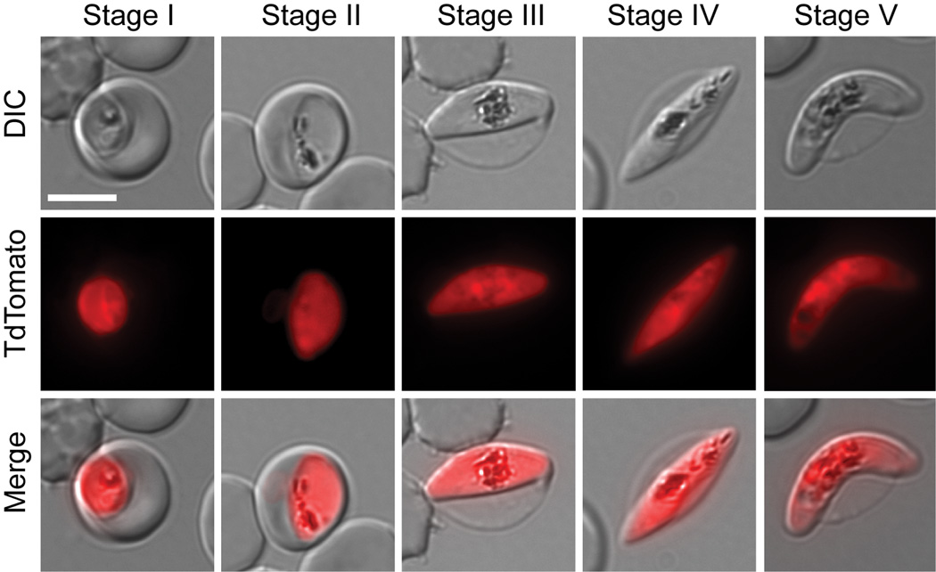
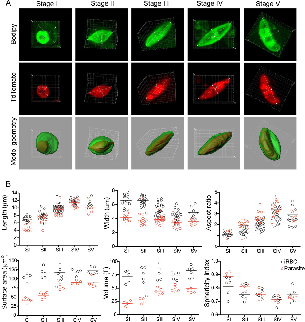
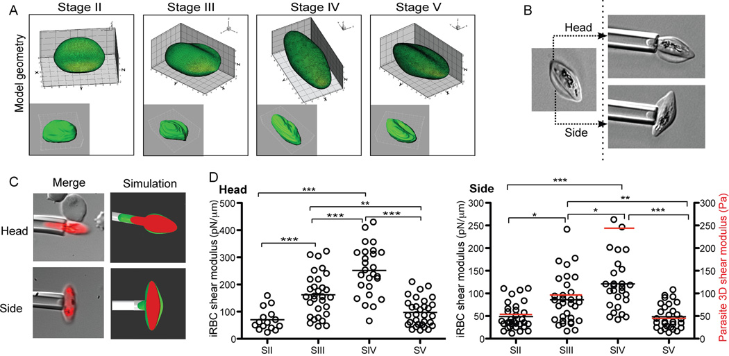
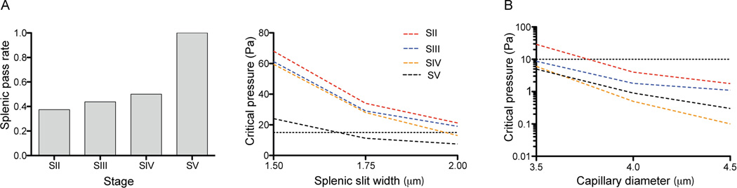
Gametocyte maturation in Plasmodium falciparum is a critical step in the transmission of malaria. While the majority of parasites proliferate asexually in red blood cells, a small fraction of parasites undergo sexual conversion and mature over 2 weeks to become competent for transmission to a mosquito vector. Immature gametocytes sequester in deep tissues while mature stages must be able to circulate, pass the spleen and present themselves to the mosquito vector in order to complete transmission. Sequestration of asexual red blood cell stage parasites has been investigated in great detail. These studies have demonstrated that induction of cytoadherence properties through specific receptor-ligand interactions coincides with a significant increase in host cell stiffness. In contrast, the adherence and biophysical properties of gametocyte-infected red blood cells have not been studied systematically. Utilizing a transgenic line for 3D live imaging, in vitro capillary assays and 3D finite element whole cell modelling, we studied the role of cellular deformability in determining the circulatory characteristics of gametocytes. Our analysis shows that the red blood cell deformability of immature gametocytes displays an overall decrease followed by rapid restoration in mature gametocytes. Intriguingly, simulations suggest that along with deformability variations, the morphological changes of the parasite may play an important role in tissue distribution in vivo. Taken together, we present a model, which suggests that mature but not immature gametocytes circulate in the peripheral blood for uptake in the mosquito blood meal and transmission to another human host thus ensuring long-term survival of the parasite.


Figures

Figure 1
Figure 1. Gametocyte development in the transgenic 164/TdTomato line
Shown are representative cells for each developmental stage as defined previously by Hawking et al (Hawking et al., 1971) and Sinden (Sinden, 1982). Stage I emerges 30 hours post invasion (pi). It is indistinguishable from the young asexual trophozoite by light microscopy. We defined Stage I parasite by shape and the presence of TdTomato fluorescence. Stage II emerges 48 –72 hours pi. We defined early Stage II parasites by the oat grain like morphology and later Stage II by the D-shaped morphology. Stage III emerges 3–5 days pi. We defined Stage III parasites by the relative elongation/flattened D-shape compared to late Stage II. Later Stage III parasites were defined by the presence of lightly pointed ends. Stage IV emerges 6–8 days pi. These parasites were defined by the spindle shape and axial symmetry with two pointed ends. Stage V emerges 10–14 days pi. This stage is mainly defined by the crescent female morphology, while the male Stage V gametocyte requires additional markers for unambiguous staging. Since male Stage V gametocytes only make up 15–20% of all Stage V gametocytes, we defined Stage V by the easily recognizable crescent shape of the female Stage V. For each cell an epifluorescence image with TdTomato distribution, differential interference contrast (DIC) and a merge of the two are shown. Images were collected at ambient temperature with a Zeiss Axioscope microscope using a 100×/1.4 immersion oil lens, and captured using a Hamamatsu Orca C4742-95 camera. Acquisition software Zeiss Axiovision was used.
Figure 2
Figure 2. 3D analysis and key parameters during sexual development. A. Gametocyte development in three dimensions
Shown is one representative cell per stage. Each cell is shown as a maximum projection; red (TdTom), green (Bodipy-FL5- Ceramide) and as a surface rendered merge of the 2 channels. Measurements for aspect ratios are included. Movies of the original files for each cell shown are available as supplementary material. Images were taken using an Olympus FV1000 confocal microscope equipped with a 100×/1.4 oil immersion lens and stacks collected using Fluoview software. Three-dimensional images were constructed using the Imaris software. B. Calculation of vital parameters. Length (x), width (y) and thickness (z) dimensions were measured from n=13 (Stage I), n=16 (Stage II), n=21 (Stage III), n=15 (Stage IV) and n=8 (Stage V). The complete data set is available as supplementary table S3.
Figure 3
Figure 3. Micropipette aspiration assays in developing gametocytes. A. Definition of cell shape
For each stage the 3D reconstruction of a representative cell was used to calculate the geometry of the iRBC as a correlate for cell shape. B/C. Schematic of capillary measurements. In order to account for the irregular shape of gametocyte-infected RBCs, the cell was aspired from side and head separately. In addition a fluorescence image of the iRBC was captured at the endpoint of the assay. D. Side and head shear modulus calculations and parasite rigidity. Shown are individual data points and mean for each stage. Average parasite rigidity is shown as a red bar in the side shear modulus graph. After ensuring that the data for each stage did not deviate severely from normality and that the assumptions for ANOVA are met, we performed a one-way ANOVA, followed by Turkey's HSD procedure to test the difference between each pair of stages. The final p-values from this test are given by *, p<0.005; **, p<0.001; ***, p<0.0005.
Figure 4
Figure 4. Simulating in vivo behavior of malaria transmission stages. A. Parametric study to simulate splenic passage of developing gametocytes
Cell shape, deformability and parasite rigidity as well as the geometry of the splenic slit was used to calculate the rate of splenic passage for each gametocyte stage (left panel). Slit width was varied from 1.5 to 2 µm and pressure drop per cell from 0 to 80 Pa. If we take the physiological pressure drop per cell as 15 Pa (dashed line), and the limiting splenic slit width as 2 µm, under these conditions the majority of immature gametocytes cannot pass. Both slit width and critical pressure are contributing to the pass rate (Right panel). B. Simulation of microcirculation. Capillary diameter was varied from 3.5 to 4.5 µm, and pressure drop per cell from 0 to 30 Pa. Under physiological flow (10 Pa pressure drop per cell, dashed line) all gametocyte stages can pass through the microcirculation at a minimal capillary diameter of 4 µm.

References

    1. Bachmann A, Esser C, Petter M, Predehl S, von Kalckreuth V, Schmiedel S, et al. Absence of erythrocyte sequestration and lack of multicopy gene family expression in Plasmodium falciparum from a splenectomized malaria patient. PLoS ONE. 2009;4:e7459. - PMC - PubMed
    1. Buchholz K, Burke TA, Williamson KC, Wiegand RC, Wirth DF, Marti M. A high-throughput screen targeting malaria transmission stages opens new avenues for drug development. The Journal of infectious diseases. 2011;203:1445–1453. - PMC - PubMed
    1. Cokelet GR. Dynamics of erythrocyte motion in filtration tests and in vivo flow. Scand J Clin Lab Invest Suppl. 1981;156:77–82. - PubMed
    1. Dao M, Lim CT, Suresh S. Mechanics of the human red blood cell deformed by optical tweezers. Journal of the Mechanics and Physics of Solids. 2003;51:2259–2280.
    1. David PH, Hommel M, Miller LH, Udeinya IJ, Oligino LD. Parasite sequestration in Plasmodium falciparum malaria: spleen and antibody modulation of cytoadherence of infected erythrocytes. Proceedings of the National Academy of Sciences of the United States of America. 1983;80:5075–5079. - PMC - PubMed

Publication types