. 2012 May 4;287(19):15896-905.
doi: 10.1074/jbc.M111.336149. Epub 2012 Mar 14.

Six-transmembrane topology for Golgi anti-apoptotic protein (GAAP) and Bax inhibitor 1 (BI-1) provides model for the transmembrane Bax inhibitor-containing motif (TMBIM) family

Affiliations

Six-transmembrane topology for Golgi anti-apoptotic protein (GAAP) and Bax inhibitor 1 (BI-1) provides model for the transmembrane Bax inhibitor-containing motif (TMBIM) family

Guia Carrara et al. J Biol Chem. .

Abstract

The Golgi anti-apoptotic protein (GAAP) is a hydrophobic Golgi protein that regulates intracellular calcium fluxes and apoptosis. GAAP is highly conserved throughout eukaryotes and some strains of vaccinia virus (VACV) and camelpox virus. Based on sequence, phylogeny, and hydrophobicity, GAAPs were classified within the transmembrane Bax inhibitor-containing motif (TMBIM) family. TMBIM members are anti-apoptotic and were predicted to have seven-transmembrane domains (TMDs). However, topology prediction programs are inconsistent and predicted that GAAP and other TMBIM members have six or seven TMDs. To address this discrepancy, we mapped the transmembrane topology of viral (vGAAP) and human (hGAAP), as well as Bax inhibitor (BI-1). Data presented show a six-, not seven-, transmembrane topology for vGAAP with a putative reentrant loop at the C terminus and both termini located in the cytosol. We find that this topology is also conserved in hGAAP and BI-1. This places the charged C terminus in the cytosol, and mutation of these charged residues in hGAAP ablated its anti-apoptotic function. Given the highly conserved hydrophobicity profile within the TMBIM family and recent phylogenetic data indicating that a GAAP-like protein may have been the ancestral progenitor of a subset of the TMBIM family, we propose that this vGAAP topology may be used as a model for the remainder of the TMBIM family of proteins. The topology described provides valuable information on the structure and function of an important but poorly understood family of proteins.


Figures

FIGURE 1.
FIGURE 1.
Potential membrane topologies of members of the GAAP family. A, amino acid sequence of VACV strain Evans GAAP and TMDs predicted by TMHMM 2.0 (blue), MEMSAT-SVM (red), TopPred 0.01(green) and SOSUI (orange). B, schematic illustration of hGAAP and BI-1 transmembrane regions (TM1–TM7) predicted by MEMSAT-SMV (black) and an additional transmembrane region predicted by MEMSAT3 (gray). Positions of individual HA tags insertions into VACV GAAP and BI-1 are indicated (black triangles).
FIGURE 2.
FIGURE 2.
Antibody probing of control proteins of known topology. A, HeLa cells transfected with plasmids expressing control Golgi resident proteins of known topology, GalT-GFP, and GRASP65-GFP (21). The orientations of fluorescent tags were verified based on their accessibility with GFP antibody following complete or cell surface permeabilization with Triton X-100 or digitonin, respectively. Scale bar, 20 μm. B, schematic representation of GalT-GFP and GRASP65-GFP topology on the Golgi membrane. C, schematic representation of selective permeabilization assay used to determine the topology of epitopes based on their accessibility with corresponding antibody. Following treatment with 20 μm digitonin, only cytosolic-orientated epitope tags (red) are accessible and stained by antibody, whereas complete permeabilization with 0.1% Triton X-100 allows detection of epitope tags regardless of their orientation. IN and OUT refer to the epitope being located on the luminal or cytoplasmic side of organelle membranes, respectively. Conditions for digitonin treatment were optimized as described by Lorenz et al. (22). D, efficiency of digitonin treatment in topology assays addressed in cells expressing control alleles GRASP65-GFP and GalT-GFP. Data are presented as the mean (± S.E.; error bars) percentage of cells positive (black) or negative (gray) for GFP antibody staining within the transfected cell population. Each experiment was carried out in triplicate. Statistical analyses were carried out using unpaired Student's t test; ***, p < 0.0005.
FIGURE 3.
FIGURE 3.
Expression of alleles. HeLa cells were either mock transfected (Mock T) or transfected with plasmids expressing different vGAAP, hGAAP, and BI-1 alleles or with plasmids expressing control proteins, GalT-GFP, and GRASP65-GFP, and cell extracts were prepared 16 h later and analyzed by immunoblotting. Allele V5-hGAAP was expressed and harvested from a HeLa cell line. The alleles tested were vGAAP-YFP, hGAAP-YFP, CFP-BI-1, BI-1-CFP, vGAAP-YFP containing a HA tag inserted between putative TMD 1–2, 2–3, 3–4, 4–5, 5–6, or 6–7, vGAAP-HA, hGAAP-HA, and V5-hGAAP.
FIGURE 4.
FIGURE 4.
N-terminal topology of members of the TMBIM family. A, membranes of HeLa cells expressing vGAAP-HA, hGAAP-HA, and N-terminally V5-tagged hGAAP were selectively or completely permeabilized with digitonin (+ Digitonin) or Triton X-100 (+ Triton), respectively. The GAAP N terminus was probed using the N terminus-specific anti-GAAP antiserum (1), and the V5 tag was probed with anti-v5 antibody. B, fluorescent protein fused at the N terminus of BI-1 was probed with cross-reactive GFP antibody (α-FP) following selective or complete permeabilization with digitonin or Triton X-100, respectively. Scale bar, 20 μm.
FIGURE 5.
FIGURE 5.
C-terminal topology of members of the TMBIM family. HeLa cells transfected with plasmids expressing vGAAP, hGAAP, or BI-1 fused at the C terminus with fluorescent or HA tags, were selectively or completely permeabilized with digitonin or Triton X-100, respectively. Tags were probed with antibody to fluorescent protein (α-FP) or to HA (α-HA). Scale bar, 20 μm.
FIGURE 6.
FIGURE 6.
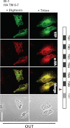
Topology of vGAAP intermembrane loops. HeLa cells were transfected with plasmids expressing vGAAP-YFP containing an HA tag (red triangles) between putative TMDs 1–2, 2–3, or 3–4. Cells were selectively permeabilized with Triton X-100 or digitonin and were probed with antibody to HA (α-HA). IN and OUT refer to the epitope being located on the luminal or cytoplasmic side of organelle membranes, respectively. Scale bar, 20 μm.
FIGURE 7.
FIGURE 7.
Topology of vGAAP intermembrane loops. HeLa cells were transfected with plasmids expressing vGAAP-YFP containing an HA tag (red triangles) between predicted TMDs 4–5, 5–6, or 6–7. Cells were selectively permeabilized with Triton X-100 or digitonin and were probed with HA antibody (α-HA). Scale bar, 20 μm.
FIGURE 8.
FIGURE 8.
Location of loop between BI-1 putative TMD 6 and 7. HeLa cells were transfected with plasmids expressing BI-1-YFP containing a HA tag (red triangle) inserted between putative TMD 6 and 7. Cells were selectively permeabilized with Triton X-100 or digitonin, and were probed with HA antibody (α-HA). Scale bar, 20 μm.
FIGURE 9.
FIGURE 9.
Proposed reentrant loop remains associated with cellular membranes. HeLa cells were transfected with plasmids expressing vGAAP-HA, vGAAP TMD 7-HA truncation, and free YFP. Cells were either fixed with paraformaldehyde and treated with 0.1% Triton X-100, or permeabilized with digitonin followed by fixation. In the latter case, membrane-free proteins were washed out, and membrane-associated proteins were probed with antibody to HA (α-HA). Scale bar, 20 μm.
FIGURE 10.
FIGURE 10.
Series of charged residues at hGAAP C terminus are required for inhibition of Fas, cisplatin, and doxorubicin-induced apoptosis. A, amino acid sequence of the human BI-1 charged C terminus required for anti-apoptotic function is underlined. Conserved residues in the hGAAP C terminus were mutated to alanine. B, expression of Neo, hGAAP, and hGAAP Cmut U2OS cell lines was immunoblotted using HA antibody. C, confocal microscopy of Neo, hGAAP, and hGAAP Cmut U2OS cell lines probed with HA antibody is shown. Scale bar, 20 μm. D–F, Neo, hGAAP, and hGAAP Cmut U2OS cell lines were mock-treated or treated with 10 μg/ml cisplatin for 24 h (D), 3 μm doxorubicin for 48 h (E), or 10 μg/ml cycloheximide alone or together with 500 ng/ml anti-Fas antibody for 24 h (F). Cells were analyzed using flow cytometry. Data are presented as the mean (± S.D. (error bars)) percentage of sub-G1 cells of a representative set of results from three independent experiments, each carried out in triplicate. Statistical analyses were carried out using unpaired Student's t test; *, p < 0.05; **, p < 0.005; ***, p < 0.0005; ****, p < 0.00005.
FIGURE 11.
FIGURE 11.
Membrane topology model of GAAPs and BI-1. The six-TMD model of vGAAP with a cytosolic orientation of both N and C termini is conserved in vGAAP, hGAAP, and BI-1. A putative reentrant loop (7) is shown with an alternative cytosolic localization (dashed line). The location of tags used in this study (v5, HA, YFP) which were used to demonstrate the TMDs 1–6, and the N-terminal peptide targeted by the GAAP antibody (arrowhead) are annotated.

References

    1. Gubser C., Bergamaschi D., Hollinshead M., Lu X., van Kuppeveld F. J., Smith G. L. (2007) A new inhibitor of apoptosis from Vaccinia virus and eukaryotes. PLoS Pathog. 3, e17. - PMC - PubMed
    1. Gubser C., Smith G. L. (2002) The sequence of camelpox virus shows it is most closely related to Variola virus, the cause of smallpox. J. Gen. Virol. 83, 855–872 - PubMed
    1. Hu L., Smith T. F., Goldberger G. (2009) LFG: a candidate apoptosis regulatory gene family. Apoptosis 14, 1255–1265 - PubMed
    1. Alcamí A., Smith G. L. (1995) Vaccinia, cowpox, and camelpox viruses encode soluble γ-interferon receptors with novel broad species specificity. J. Virol. 69, 4633–4639 - PMC - PubMed
    1. Nuara A. A., Walter L. J., Logsdon N. J., Yoon S. I., Jones B. C., Schriewer J. M., Buller R. M., Walter M. R. (2008) Structure and mechanism of IFN-γ antagonism by an orthopoxvirus IFN-γ-binding protein. Proc. Natl. Acad. Sci. U.S.A. 105, 1861–1866 - PMC - PubMed

Publication types

MeSH terms