

Visualizing molecular juggling within a B12-dependent methyltransferase complex
- PMID: 22419154
- PMCID: PMC3326194
- DOI: 10.1038/nature10916
Visualizing molecular juggling within a B12-dependent methyltransferase complex
Abstract
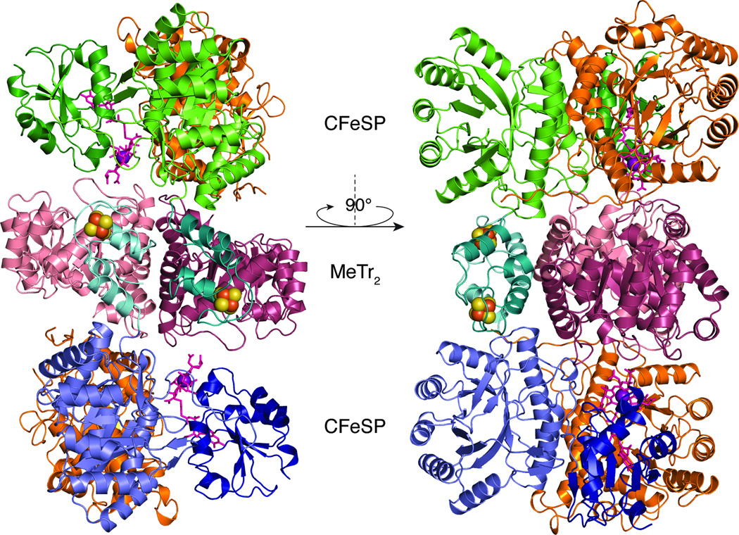
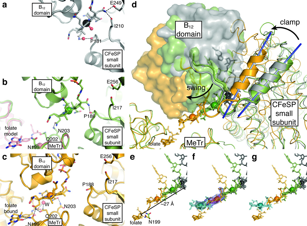
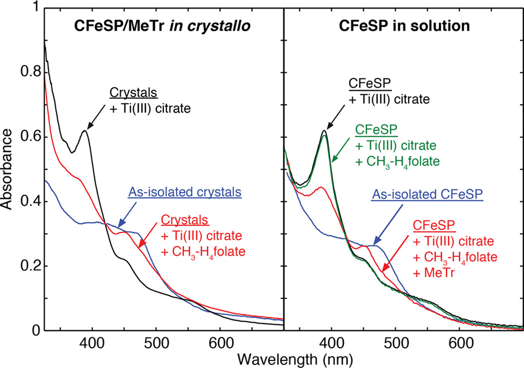
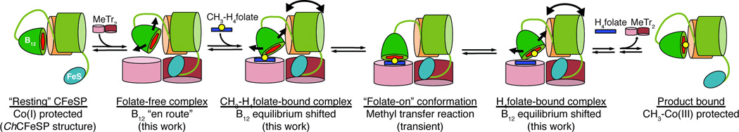
Derivatives of vitamin B(12) are used in methyl group transfer in biological processes as diverse as methionine synthesis in humans and CO(2) fixation in acetogenic bacteria. This seemingly straightforward reaction requires large, multimodular enzyme complexes that adopt multiple conformations to alternately activate, protect and perform catalysis on the reactive B(12) cofactor. Crystal structures determined thus far have provided structural information for only fragments of these complexes, inspiring speculation about the overall protein assembly and conformational movements inherent to activity. Here we present X-ray crystal structures of a complete 220 kDa complex that contains all enzymes responsible for B(12)-dependent methyl transfer, namely the corrinoid iron-sulphur protein and its methyltransferase from the model acetogen Moorella thermoacetica. These structures provide the first three-dimensional depiction of all protein modules required for the activation, protection and catalytic steps of B(12)-dependent methyl transfer. In addition, the structures capture B(12) at multiple locations between its 'resting' and catalytic positions, allowing visualization of the dramatic protein rearrangements that enable methyl transfer and identification of the trajectory for B(12) movement within the large enzyme scaffold. The structures are also presented alongside in crystallo spectroscopic data, which confirm enzymatic activity within crystals and demonstrate the largest known conformational movements of proteins in a crystalline state. Taken together, this work provides a model for the molecular juggling that accompanies turnover and helps explain why such an elaborate protein framework is required for such a simple, yet biologically essential reaction.
Figures
References
-
- Matthews RG. Cobalamin-Dependent Methyltransferases. Acc. Chem. Res. 2001;34:681–689. - PubMed
-
- Banerjee RB, Ragsdale SW. The Many Faces of Vitamin B12: Catalysis by Cobalamin-Dependent Enzymes. Annu. Rev. Biochem. 2003;72:209–247. - PubMed
-
- Drennan CL, Huang S, Drummond JT, Matthews RG, Ludwig ML. How a Protein Binds B12: A 3.0 Å X-ray Structure of B12-Binding Domains of Methionine Sythase. Science. 1994;266:1669–1674. - PubMed
-
- Dixon MM, Huang S, Matthews RG, Ludwig ML. The Structure of the C-terminal Domain of Methionine Synthase: Presenting S-adenosylmethionine for Reductive Methylation of B12. Structure. 1996;4:1263–1275. - PubMed
Publication types
MeSH terms
Substances
Associated data
- Actions
- Actions
- Actions
Grants and funding
LinkOut - more resources
Full Text Sources
Other Literature Sources
Molecular Biology Databases
