

Case Reports
doi: 10.1002/ajmg.a.35261.
Epub 2012 Mar 14.
Early-onset osteoarthritis in Ehlers-Danlos syndrome type VIII
Affiliations
- PMID: 22419391
- PMCID: PMC3314117
- DOI: 10.1002/ajmg.a.35261
Item in Clipboard
Case Reports
Early-onset osteoarthritis in Ehlers-Danlos syndrome type VIII
Am J Med Genet A.
2012 Apr.
No abstract available
Figures
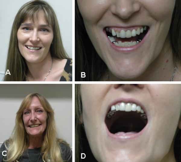
Facial and dental appearance of family with EDS type VIII. A, B, and D – proposita; C – her mother. Note long nose with narrow root and prominent tip that have been reported in patients with this type of EDS. Patients wear dentures.
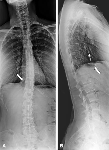
Anterior-posterior and lateral spine radiographs. Note moderate S-shaped scoliosis of the spine with maximum point at the thoracic-lumbar junction [A] and decreased bone density as observed on conventional radiography with apparent increased density of the vertebral endplates [B]. In addition, an abnormal pre/paravertebral air-fluid-level collection [arrows] is demonstrated on both planes indicative of a gastro-esophageal hernia [A, B].
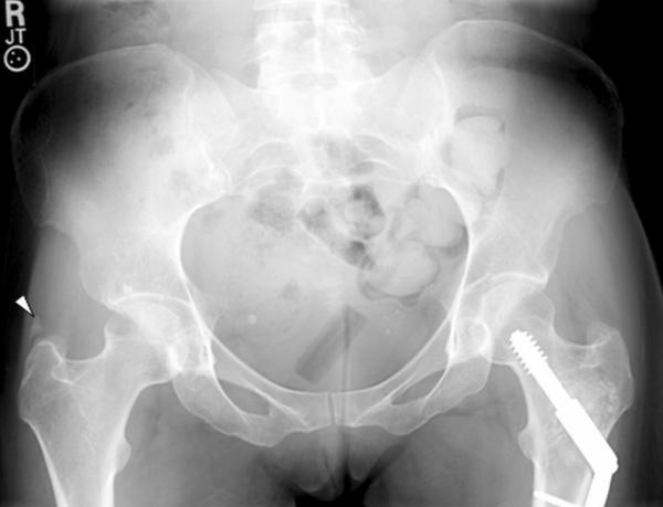
Pelvic radiographs. Anterior-posterior radiograph of the pelvis. Note normal joint spaces of the hip joints on both sides without obvious degeneration. On the right side, adjacent to the greater trochanter there is an abnormal ossicle [arrow] suggesting tendinopathy of the gluteal muscles. The left femur demonstrates postsurgical changes with osteosynthetic treatment of the femoral neck.
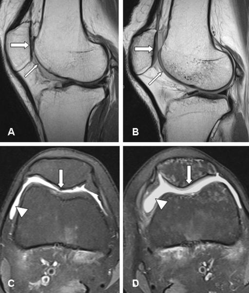
MR imaging of the knee joints. Sagittal [A, B] and transverse [C, D] images of the right [A, C] and left [B, D] knee joint, respectively. On the right side, absence of the articular cartilage [thin arrow] of the femoral joint surface [A] and marked defects and thinning of the retro-patellar cartilage [thick arrow]; whereas the cartilaginous layers [arrows] of the left knee are only mildly thinned [B]. The transverse images feature joint effusion on both sides [arrowheads] [C, D], and again distinct cartilage damage of the right retro-patellar surface [arrow] [C], and slight cartilaginous changes left-sided [arrow] [D].
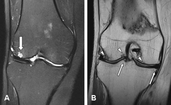
Coronal MR imaging of the right knee joint. The lateral joint compartment demonstrates a distinct cyst [arrow] of the subchondral bone [A], and multiple surface and subchondral irregularities [arrowheads], as well as osteophytic spurs [arrows] [B], which are overall the result of marked degeneration.
References
-
- Biro F, Gewanter HL, Baum J. The hypermobility syndrome. Pediatrics. 1983;72:701–706. - PubMed
-
- Castori M, Camerota F, Celletti C, Danese C, Santilli V, Saraceni VM, Grammatico P. Natural history and manifestations of the hypermobility type Ehlers–Danlos syndrome: A pilot study on 21 patients. Am J Med Genet. 2010;152:556–564. - PubMed
-
- Karachalios T, Zibis A, Papanagiotou P, Karantanas AH, Malizos KN, Roidis N. MR imaging findings in early osteoarthritis of the knee. Eur J Radiol. 2004;50:225–230. - PubMed
-
- Karrer S, Landthaler M, Schmalz G. Ehlers–Danlos type VIII. Review of the literature. Clin Oral Investig. 2000;4:66–69. - PubMed
-
- Moore MM, Votava JM, Orlow SJ, Schaffer JV. Ehlers–Danlos syndrome type VIII: Periodontitis, easy bruising, marfanoid habitus, and distinctive facies. J Am Acad Dermatol. 2006;55:S41–S45. - PubMed
Publication types
MeSH terms
Grants and funding
LinkOut - more resources
Full Text Sources
Medical
