. 2012 May;97(5):E719-30.
doi: 10.1210/jc.2011-3251. Epub 2012 Mar 14.

Progesterone receptor-A and -B have opposite effects on proinflammatory gene expression in human myometrial cells: implications for progesterone actions in human pregnancy and parturition

Affiliations

Progesterone receptor-A and -B have opposite effects on proinflammatory gene expression in human myometrial cells: implications for progesterone actions in human pregnancy and parturition

Huiqing Tan et al. J Clin Endocrinol Metab. 2012 May.

Abstract

Context: Progesterone promotes uterine relaxation during pregnancy and its withdrawal induces labor. Progesterone withdrawal in human parturition is mediated in part by changes in the relative levels of the nuclear progesterone receptor isoforms, PR-A and PR-B, in myometrial cells. Parturition also involves myometrial inflammation; however, the functional link between nuclear PR-mediated progesterone actions and inflammation in human myometrial cells is unclear.

Objective: Our objective was to determine how PR-A and PR-B regulate progesterone action in human myometrial cells and specifically the expression of genes encoding contraction-associated proteins and proinflammatory mediators.

Design: Effects of PR-A and PR-B on the capacity for progesterone to modulate gene expression was determined using an immortalized human myometrial cell line stably transfected with inducible PR-A and PR-B expression transgenes and conditioned to express various PR-A and PR-B levels. Gene expression was assessed by genome wide transcriptome analysis, quantitative RT-PCR and immunoblotting.

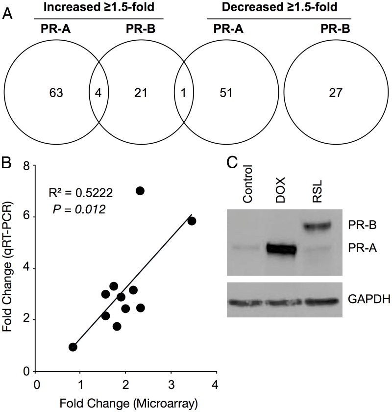
Results: PR-A and PR-B were each transcriptionally active in response to progesterone and affected the expression of distinct gene cohorts. The capacity for progesterone to affect gene expression was dependent on the PR-A to PR-B ratio. This was especially apparent for the expression of proinflammatory genes. Progesterone decreased proinflammatory gene expression when the PR-A to PR-B ratio favored PR-B and increased proinflammatory gene expression when the ratio favored PR-A. Progesterone via PR-B increased expression of inhibitor-κBα, a repressor of the nuclear factor-κB transcription factor, and inhibited basal and lipopolysaccharide-induced proinflammatory gene expression. Both of those PR-B-mediated effects were inhibited by PR-A.

Conclusions: Our data suggest that during most of human pregnancy, when myometrial cells are PR-B dominant, progesterone promotes myometrial quiescence through PR-B-mediated antiinflammatory actions. At parturition, the rise in PR-A expression promotes labor by inhibiting the antiinflammatory actions of PR-B and stimulating proinflammatory gene expression in response to progesterone.


Figures

Fig. 1.
Fig. 1.
Inducible expression of functional PR-A and PR-B from stably incorporated TetON-Advanced (PR-A) and RheoSwitch (PR-B) transgenes in hTERT-HMA/B cells. A, Whole-cell lysates of hTERT-HMA/B cells treated with varying concentrations of DOX and RSL for 48 h assessed by immunoblot analysis for nPR (5 min exposure) and glyceraldehyde 3-phosphate dehydrogenase (GAPDH) (30 sec exposure). B, nPR immunolocalization (PgR1294 antibody; bar, 100 μm) in hTERT-HMA/B cells exposed to vehicle or DOX (10 ng/ml) for 24 h. Consistent with the immunoblot outcomes (far right lane), the cells expressed low levels of nPR under basal conditions. DOX induced expression of PR-A (indicated by grey intensity) in all cells. Identical outcomes were obtained with cells exposed to RSL (data not shown).
Fig. 2.
Fig. 2.
Effects of PR-A and PR-B on gene expression in hTERT-HMA/B cells. A, Venn diagram showing numbers of genes affected (≥1.5-fold change compared with nPR expressing cells in the absence of progesterone) by progesterone (100 nm for 6h) via PR-A and PR-B. Only annotated genes encoding known proteins were included in the analysis. B, Correlation analysis (Pearson's correlation analysis) between microarray and qRT-PCR outcomes for 11 different genes affected by PR-A and PR-B. C, nPR immunoblot analysis of total cell lysates from hTERT-HMA/B cells used for the microarray analysis. Cells were exposed to DOX (50 ng/ml) and RSL (300 nm) for 24 h. GAPDH, Glyceraldehyde 3-phosphate dehydrogenase.
Fig. 3.
Fig. 3.
Effects of the PR-A to PR-B ratio on progesterone-induced gene expression in hTERT-HMA/B cells. A, Effect of PR-A to PR-B ratio levels on basal and progesterone-induced expression of PTGS2, IL8, IL1B, and GJA1 in hTERT-HMA/B cells conditioned with DOX and RSL to be in PR-B- or PR-A-dominant states (indicated by the immunoblot). Cells were exposed to progesterone or vehicle for 16 h and then various concentrations of DOX and RSL for 12 h to modify the PR-A and PR-B levels. (Data are relative to abundance of mRNA encoding α-actin in each experimental condition; mean ± se; n = 3; *, P < 0.05.) B, Immunoblot analysis of PTGS2 and IL-8 levels in hTERT-HMA/B cells exposed to RSL and various levels of DOX ± progesterone (100 nm). Data are representative of triplicate experiments.
Fig. 4.
Fig. 4.
Effects of PR-A and PR-B on progesterone (100 nm, 24 h)-induced expression of NFKB1A and FKBP5 in hTERT-HMA/B cells. Progesterone via PR-B increased NFKB1A and FKBP5 expression. PR-A inhibited the effects of PR-B on NFKB1A expression in a dose-dependent manner but did not affect PR-B-mediated expression of FKBP5 (data are relative to abundance of mRNA encoding α-actin in each experimental condition; mean ± se; n = 3; *, P < 0.05).
Fig. 5.
Fig. 5.
Effects of progesterone (P4) and the PR-A to PR-B ratio on basal and LPS-induced expression of PTGS2 in hTERT-HMA/B cells. As expected, LPS increased PTGS2 expression. In PR-B-dominant cells, progesterone inhibited basal and LPS-induced PTGS2 expression. In PR-A-dominant cells, progesterone increased basal PTGS2 expression and did not affect LPS-induced PTGS2 expression. Cells were exposed to vehicle (Veh) or RSL and DOX ± P4 (100 nm) for 24 h ± LPS (1 μg/ml; final 6 h). Immunoblot: nPR expression achieved by DOX and RSL treatment (data are relative to abundance of mRNA encoding α-actin in each experimental condition; mean ± se; n = 3; *, P < 0.05). GAPDH, Glyceraldehyde 3-phosphate dehydrogenase.
Fig. 6.
Fig. 6.
Working model. Progesterone via PR-B inhibits proinflammatory gene expression and suppresses responsiveness of myometrial cells to proinflammatory/prolabor stimuli by stimulating expression of NFKB1A, which inhibits NF-κB activity. PR-A blocks the antiinflammatory actions of progesterone mediated by PR-B (e.g. by blocking PR-B induction of NFKB1A expression) and directly increases proinflammatory gene expression (indicated by PTGS2), leading to a proinflammatory state within the myometrial compartment. PR-A expression is increased by PGF whose levels increase in response to prolabor stimuli. As pregnancy progresses, prolabor stimuli (e.g. uterine distention, intrauterine infection, and fetal membrane PG production) increase local PGF levels within the gestational tissues, leading to increased myometrial cell PR-A expression and a gradual increase in the PR-A to PR-B ratio in myometrial cells. A PR-A to PR-B threshold exists above which PR-A represses the antiinflammatory activity of PR-B and begins to mediate proinflammatory effects of progesterone. At this point a positive-feedback proinflammatory loop develops within the myometrium whereby increased production of PGF by myometrial cells (due to progesterone/PR-A mediated increased PTGS2 expression) exerts an autocrine/paracrine effect that further augments PR-A expression that further increases the inflammatory state leading to a further increase in PGF. Eventually the PGF levels increase to a point whereby the hormone exerts potent uterotonic actions to induce labor. FP, PGF receptor; PGR, progesterone receptor.

References

    1. Corner GW. 1946. The hormones in human reproduction. London: Princeton University Press
    1. Csapo A. 1956. Progesterone block. Am J Anat 98:273–291 - PubMed
    1. Sanborn BM. 2000. Relationship of ion channel activity to control of myometrial calcium. J Soc Gynecol Investig 7:4–11 - PubMed
    1. Sanborn BM, Yue C, Wang W, Dodge KL. 1998. G protein signalling pathways in myometrium: affecting the balance between contraction and relaxation. Rev Reprod 3:196–205 - PubMed
    1. Sanborn BM. 1995. Ion channels and the control of myometrial electrical activity. Semin Perinatol 19:31–40 - PubMed

Publication types