. 2012 May;25(3):312-25.
doi: 10.1111/j.1755-148X.2012.00998.x.

Constitutive gray hair in mice induced by melanocyte-specific deletion of c-Myc

Affiliations

Constitutive gray hair in mice induced by melanocyte-specific deletion of c-Myc

Irina Pshenichnaya et al. Pigment Cell Melanoma Res. 2012 May.

Abstract

c-Myc is involved in the control of diverse cellular processes and implicated in the maintenance of different tissues including the neural crest. Here, we report that c-Myc is particularly important for pigment cell development and homeostasis. Targeting c-Myc specifically in the melanocyte lineage using the floxed allele of c-Myc and Tyr::Cre transgenic mice results in a congenital gray hair phenotype. The gray coat color is associated with a reduced number of functional melanocytes in the hair bulb and melanocyte stem cells in the hair bulge. Importantly, the gray phenotype does not progress with time, suggesting that maintenance of the melanocyte through the hair cycle does not involve c-Myc function. In embryos, at E13.5, c-Myc-deficient melanocyte precursors are affected in proliferation in concordance with a reduction in numbers, showing that c-Myc is required for the proper melanocyte development. Interestingly, melanocytes from c-Myc-deficient mice display elevated levels of the c-Myc paralog N-Myc. Double deletion of c-Myc and N-Myc results in nearly complete loss of the residual pigmentation, indicating that N-Myc is capable of compensating for c-Myc loss of function in melanocytes.


Figures

Figure 1
Figure 1
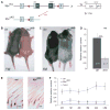
Melanocyte-specific ablation of c-Myc causes hair graying. (A) Deletion of the c-Myc gene by pigment-cell-specific expression of Cre recombinase, using mice carrying a conditional allele of c-Myc [c-Mycflox (Trumpp et al., 2001)] and the Tyr::Cre (Delmas et al., 2003) transgene. Unless specified, c-Mycflox /flox; Tyr::Cre mice are referred as MyccKO, and c-Mycflox /+; Tyr::Cre mice were used as a control (ctrl). (B, C) Mice lacking c-Myc in melanocytes show dilution of coat color, which is seen with onset of pigmentation. Mice are 1 week (B) and 1 month (C) old. (D) A significant decrease in melanin concentration was observed in the dorsal hair of MyccKO (n = 7, light gray) mice in comparison with the control animals (n = 6, dark gray) (P < 0.001). The hair samples from 1-month-old animals were analyzed. Each sample was measured at least twice. (E) H&E staining of dorsal skin section of 1-week-old animals (ctrl, left; MyccKO, right). Reduced pigmentation is seen in bulbs of hair follicles of ctrl mice. Scale bar 100 μm. (F) Melanin concentration in the dorsal hair of ctrl and MyccKO mice. Animals (on an Agouti background) were analyzed from 2 weeks (2W) to over 4 months (4M). At least two mice per experiment were analyzed.
Figure 2
Figure 2
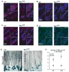
Analysis of recombination efficiency as mediated by the Tyr::Cre transgene. (A, B) c-Mycflox /flox; Tyr::Cre animals were mated to either Dct::LacZ (A, melanocyte-specific reporter line) mice to visualize melanoblasts, or Rosa26R::LacZ (B, Cre-reporter line) mice to trace recombined cells. Melanocytes were visualized as X-Gal-positive cells in E14.5 embryos. c-Mycflox /+; Tyr::Cre; R26R::LacZ or c-Mycflox /+; Tyr::Cre; Dct::LacZ mice were used as a control. Note the number of melanoblasts in MyccKO embryos is reduced and comparable in both Rosa26R::LacZ and Dct::LacZ reporter lines. Scale bar, 100 μm. (C, D) Immunostaining against LacZ and Pax3 on skin section of 1-week-old control – ctrl (C, c-Mycflox /+; Tyr::Cre; R26R::LacZ) and MyccKO (D, c-Mycflox /flox; Tyr::Cre; R26R::LacZ) mice. Double positive (LacZ+ /Pax3+) cells are detected in both the bulge (arrowhead) and bulb (arrow) compartments of the hair follicle, thus demonstrating that Cre-expressing melanoblasts and melanocytes have contributed to the post-natal melanocyte population of MyccKO mice.
Figure 3
Figure 3
c-Myc is important to maintain proper melanocyte number after birth but does not change the pattern of melanocyte-specific markers. (A–D) Immunostaining against melanocyte-specific markers in differentiated pigment cells of the bulb of the hair follicle at the beginning of anagen (A, tyrosinase; B, MITF) and in melanocyte progenitors of the bulge of the hair follicle at the beginning of anagen (C, Dct) and middle of anagen (D, Pax3). MyccKO melanocytes show the expression of pigment cell markers but fewer positive cells are detected in comparison with the control tissue. The dorsal skin of 1-week-old mice was analyzed. Scale bar, 100 μm. (E) Melanoblasts in the bulge (Bg) region of the hair follicles of 8-day-old mice were visualized by whole-mount X-gal staining of control (c-Mycflox /+; Tyr::Cre; Dct::LacZ) and conditional knockout (MyccKO: c-Mycflox /flox; Tyr::Cre; Dct::LacZ) skin. Scale bar, 100 μm. (F) Quantification of the bulge melanoblasts in the dorsal skin at 8 and 30 days after birth. Average numbers of LacZ+ cells were counted per field view in the bulge zone and normalized per hair follicle. The same areas were evaluated in the control and MyccKO mice. At least three animals per genotype were analyzed.
Figure 4
Figure 4
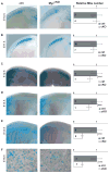
c-Myc is required for proper melanocyte development. The number of melanocyte precursors is reduced in MyccKO embryos. The Dct::LacZ reporter mouse was used to visualize the melanocytes during embryogenesis. (A–F) X-Gal staining of the control [ctrl: Tyr::Cre; Dct::LacZ (left panel)] and conditional knockout [MyccKO: c-Mycflox /flox; Tyr::Cre; Dct::LacZ (right panel)] embryos from E10.5 to E16.5. Relative melanoblast number is shown from E10.5 till E16.5. Number of cells in the control was set to one. Melanoblast number was quantified per 1 mm2 of embryo in the region between fore- and hindlimbs (E10.5–E14.5), or in a defined view of the embryo (E16.5). The number of embryos analyzed per genotype is indicated in brackets.
Figure 5
Figure 5
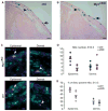
During embryogenesis, c-Myc is required to maintain a proper number of melanoblasts upon entry into the epidermis. (A–B) Representative view of melanoblast distribution in the dermis (arrowhead) and epidermis (arrow) of control, Dct::LacZ (A) and MyccKO, Dct::LacZ (B) E13.5 embryos. Transverse sections were counterstained with nuclear fast red. Scale bar, 100 μm. (C) Immunostaining against BrdU (green) and LacZ (red) shows proliferating melanoblasts in E13.5 control and MyccKO embryos. Scale bar, 25 μm. (D) The number of melanoblasts was counted in the dermis and epidermis of E13.5 control (n = 5) and MyccKO (cKO, n = 5) embryos using the Dct::LacZ reporter mouse. The number of melanoblasts per section per one lateral side of embryo is shown. The dermal melanoblast population was not affected, while the number of epidermal melanoblasts is 2.5-fold reduced upon c-Myc loss (epidermis, ctrl versus cKO – P < 0.005, while the difference in the number of dermal melanoblasts is not statistically significant). (E) The number of proliferating melanoblasts in epidermis but not dermis (see C) is reduced. The percentage of double LacZ+ /BrdU+ cells is shown in relation to total amount of LacZ+ cells in the control (n = 5, gray circles) and knockout MyccKO (n = 6, white circles) embryos (proliferation in epidermis: P < 0.02; the difference in the number of proliferating dermal melanoblasts (ctrl versus cKO) is not statistically significant).
Figure 6
Figure 6
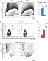
N-Myc is upregulated in c-Myc-deficient melanocytes. (A–C) FACS analysis of melanocytes in c-Myc-deficient mice (cKO: c-Mycflox /flox; Tyr::Cre). (A) SSC /FSC distribution of single cell suspension derived from c-MyccKO mice (P2). Note that the amount of granular cells (SShigh) is diminished in comparison with the control [similar to the Tyr::DT-A transgenic mouse (Figure S4A)]. (B) The number of c-kit-positive cells is decreased in c-MyccKO mice. Cells were pregated on alive CD45-negative population. (C) SSC /FSC distribution of CD45- /c-kit+ cells. The SShigh population of differentiated cells (gated in pink) is severely affected in c-MyccKO skin. Note that numbers represent a percentage of the parental population. (D, E) Melanocytes of c-MyccKO mice show a more than 8-time reduction in the level of c-Myc mRNA (P < 0.05) and a more than two times upregulation of N-Myc mRNA levels (P < 0.05). CD45- /c-kit+ /SShigh cells were FACS-sorted from c-MyccKO and control mice (P2), and the relative level of c-Myc and N-Myc mRNA was analyzed by qPCR (control, left; c-MyccKO, right).
Figure 7
Figure 7
Deletion of both c-Myc and N-Myc in melanocytes leads to further reduction in embryonic melanoblast number and severe coat color dilution after birth. (A) A N-Myc and c-Myc double knockout in the melanocyte lineage leads to a further dilution of coat pigmentation. Coat colors of the control, cKO (c-Myc conditional knockout: c-Mycflox /flox; Tyr::Cre), and dKO (c-Myc and N-Myc double knockout: c-Mycflox /flox; N-Mycflox /flox; Tyr::Cre) mice are shown at 1 week and 2 months of age. (B) Melanin concentration is decreased in the dorsal hair of 2-month-old mice, and almost no melanin is left in the double knock-out animals (dKO; P < 0.0001). (C) Almost no pigment cells can be detected in the dorsal hair follicles of dKO skin (1-week-old mice, nuclear fast red staining). (D) The Dct::LacZ reporter mouse was used to visualize the melanoblasts during embryogenesis (E14.5). Left panel: control c-Mycflox /+; Tyr::Cre; Dct::LacZ; middle panel: cKO: c-mycflox /flox;Tyr::Cre; Dct::LacZ; right panel: dKO c-Mycflox /flox; N-Mycflox /flox; Tyr::Cre; Dct::LacZ. (E) Reduction in melanoblast number at E14.5. Number of cells in the control was set to one (ctrl, n = 8 embryos; cKO, n = 6; dKO, n = 5; P < 0.0001).

References

    1. Adameyko I, Lallemend F, Aquino JB, et al. Schwann cell precursors from nerve innervation are a cellular origin of melanocytes in skin. Cell. 2009;139:366–379. - PubMed
    1. Aydin IT, Beermann F. A MART-1::Cre transgenic line induces recombination in melanocytes and RPE. Genesis. 2011;49:403–409. - PubMed
    1. Barrett J, Birrer MJ, Kato GJ, Dosaka-Akita H, Dang CV. Activation domains of L-Myc and c-Myc determine their transforming potencies in rat embryo cells. Mol Cell Biol. 1992;12:3130–3137. - PMC - PubMed
    1. Batsche E, Muchardt C, Behrens J, Hurst HC, Cremisi C. RB and c-Myc activate expression of the E-cadherin gene in epithelial cells through interaction with transcription factor AP-2. Mol Cell Biol. 1998;18:3647–3658. - PMC - PubMed
    1. Bellmeyer A, Krase J, Lindgren J, LaBonne C. The protooncogene c-myc is an essential regulator of neural crest formation in xenopus. Dev Cell. 2003;4:827–839. - PubMed

Publication types