. 2012 Jul;12(7):1708-19.
doi: 10.1111/j.1600-6143.2012.04009.x. Epub 2012 Mar 15.

The spleen is the major source of antidonor antibody-secreting cells in murine heart allograft recipients

Affiliations

The spleen is the major source of antidonor antibody-secreting cells in murine heart allograft recipients

A Sicard et al. Am J Transplant. 2012 Jul.

Abstract

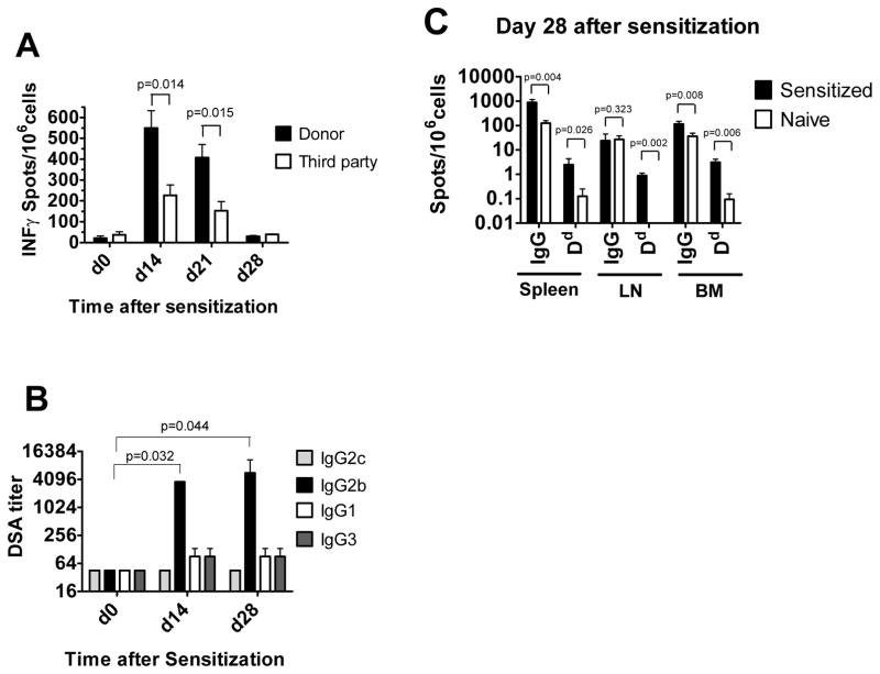
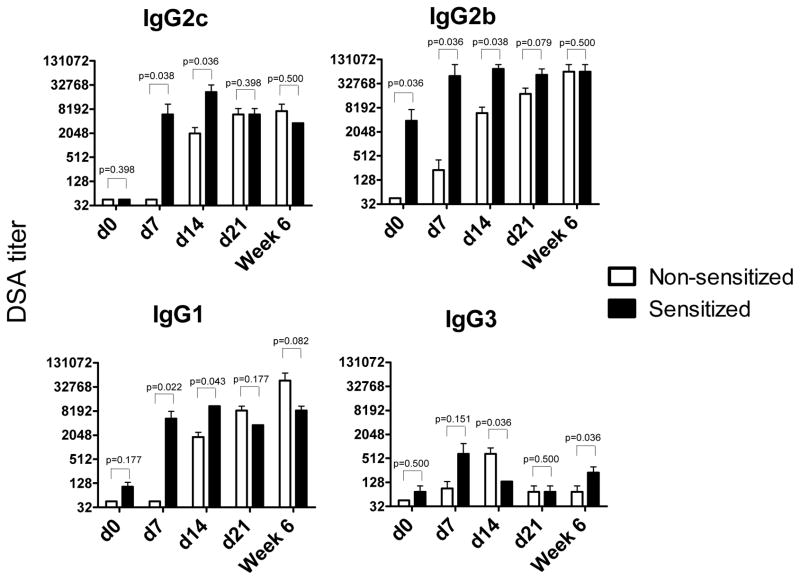
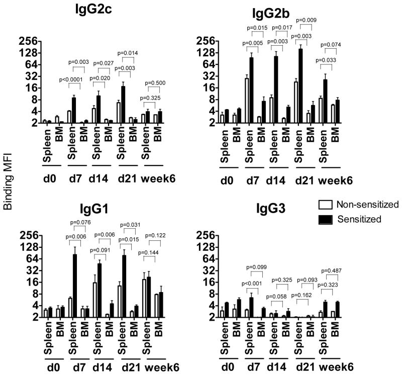
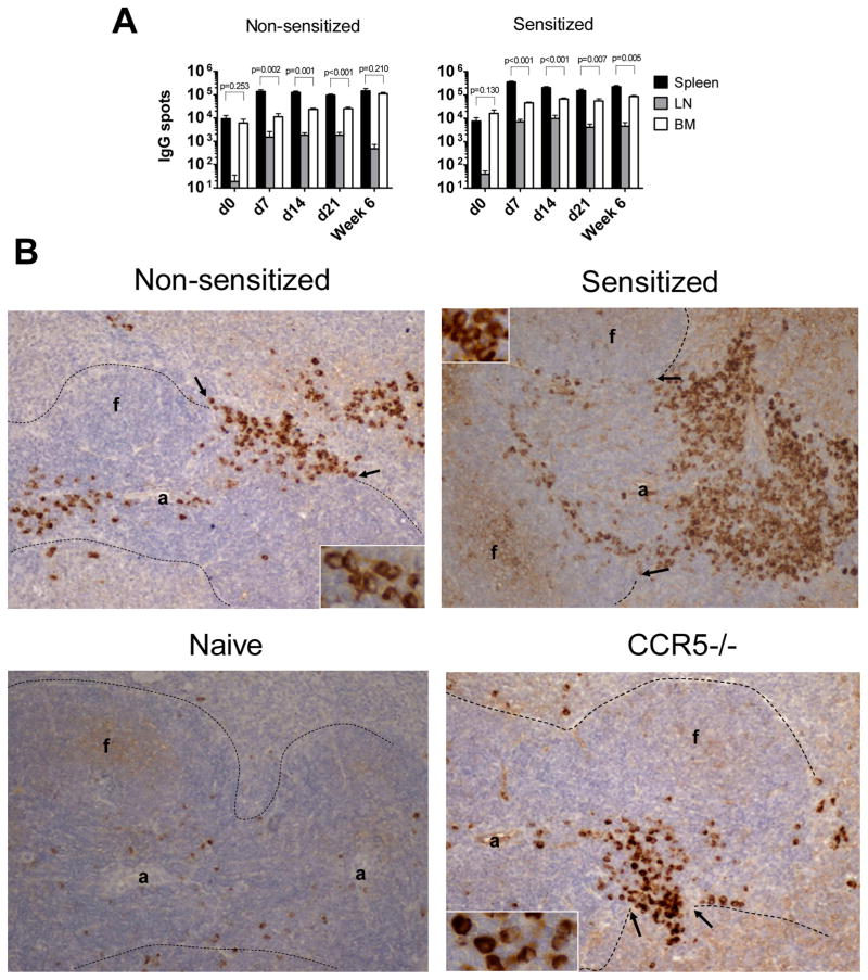
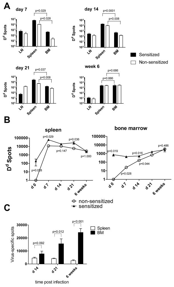
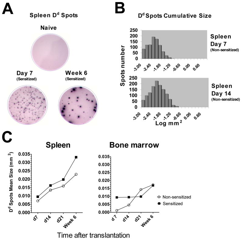
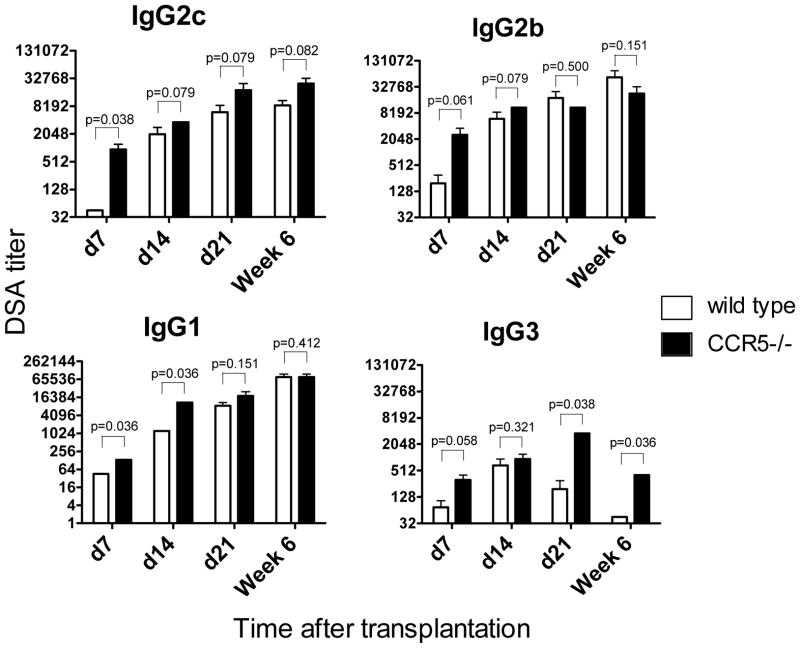
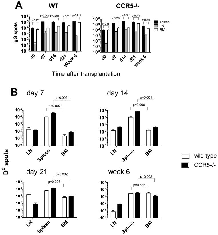
Antibody-mediated allograft rejection is an increasingly recognized problem in clinical transplantation. However, the primary location of donor-specific alloantibody (DSA)-producing cells after transplantation have not been identified. The purpose of this study was to test the contribution of allospecific antibody-secreting cells (ASCs) from different anatomical compartments in a mouse transplantation model. Fully MHC-mismatched heart allografts were transplanted into three groups of recipients: nonsensitized wild type, alloantigen-sensitized wild-type and CCR5(-/-) mice that have exaggerated alloantibody responses. We found that previous sensitization to donor alloantigens resulted in the development of antidonor alloantibody (alloAb) with accelerated kinetics. Nevertheless, the numbers of alloantibody-secreting cells and the serum titers of antidonor IgG alloantibody were equivalent in sensitized and nonsensitized recipients 6 weeks after transplantation. Regardless of recipient sensitization status, the spleen contained higher numbers of donor-reactive ASCs than bone marrow at days 7-21 after transplantation. Furthermore, individual spleen ASCs produced more antidonor IgG alloantibody than bone marrow ASCs. Taken together, our results indicate that the spleen rather than bone marrow is the major source of donor-reactive alloAb early after transplantation in both sensitized and nonsensitized recipients.


Figures

Figure 1
Figure 1. Generation of allosensitized transplant recipients
B6 mice were subcutaneously injected with BALB/c splenocytes in CFA. A. IFN γ ELISPOT assays were performed on spleen cells isolated prior to sensitization and at days 14 (n=3), 21 (n=3) and 28 (n=4) after sensitization and re-stimulated with BALB/c or third party splenocytes. B. Sera were collected prior to sensitization and on days 14 (n=4) and 28 (n=4) after sensitization, BALB/c reactive antibody titers were measured as outlined in the Methods. The titers of BALB/c reactive antibody in the serum of naïve B6 mice was < 45 for all IgG isotypes. C. IgG ELISPOT assays performed on spleen, BM and inguinal lymph node cells to enumerate the frequencies of ASCs producing total and Dd-binding IgG (n=4).
Figure 2
Figure 2. The kinetics of DSA production in sensitized versus non-sensitized heart allograft recipients
Heterotopic BALB/c heart transplants were placed into non-sensitized B6 mice or into sensitized B6 recipients four weeks after immunization with BALB/c alloantigens. Serum was collected on days 7, 14 and 21 and at six weeks post transplant, and the titers of BALB/c- and third party-reactive alloAb were determined by cell binding assay. The titers of third party-reactive alloAb in all serum samples were < 45 for IgG2c and IgG2b and < 135 for IgG1 and IgG3. The results were compared using non-parametric Mann-Whitney test. N = 4–5 mice per group at each time point.
Figure 3
Figure 3. In vitro DSA secretion by spleen and bone marrow cells
Spleen and BM cells were isolated from heart allograft recipients at 7, 14, 21 and at six weeks post transplant and cultured in vitro for 48 hours without re-stimulation. The presence of DSA was evaluated by testing the ability of the culture supernatants to bind donor thymocytes. Results are expressed as Mean Fluorescent Intensity (MFI). Spleen cells isolated from naïve B6 mice and cultured for 48 hours were used as a control. N = 4–5 mice per group at each time point.
Figure 4
Figure 4. Distribution of IgG producing ASCs within spleen, mediastinal lymph nodes and bone marrow of sensitized and non-sensitized recipients
A. Spleen, BM and mediastinal lymph nodes cells were isolated at indicated time points after transplantation or from naïve B6 mice as a control. The total numbers of IgG-secreting cells per each compartment were determined by ELISPOT assay. N = 4–5 mice per group at each time point. B. Immunohistochemical staining of recipient spleen sections for IgG secreting cells at day 7 post transplant. Spleen tissues were excised, fixed and embedded in paraffin. 5μm sections were prepared and stained with anti-mouse IgG antibodies. Dotted lines indicate the border between white and red pulp. Arrows point to bridging channels with clusters of IgG+ ASCs; “a” = arteriole, “f” = follicle. Original magnification 20x, insets 60x.
Figure 5
Figure 5. Distribution of antigen-specific IgG producing ASCs in heart allograft recipients is distinct from that in virus-infected mice
A–B. Heterotopic BALB/c heart transplants were placed into non-sensitized B6 mice or into sensitized B6 recipients four weeks after immunization with BALB/c alloantigens. Spleen, BM and mediastinal lymph nodes cells were isolated at indicated time points after transplantation. The total numbers per compartment of cells secreting Dd-binding IgG were determined by ELISPOT assay (A). The kinetics of Dd-reactive ASC frequencies in the spleen and in the BM of sensitized versus non-sensitized recipients (B). C. B6 mice were intraperitoneally injected with 5 × 10⁶ PFU of the mouse hepatitis virus strain JHMV. The numbers of virus-specific ASCs were determined by ELISPOT assay at indicated time points after infection. N= 4–5 mice per group at each time point.
Figure 6
Figure 6. Secretion of donor MHC binding IgG by individual ASCs
A–B. ELISPOT well images and cumulative spot size histograms illustrate increasing production of allo-reactive antibody by individual cells. C. The kinetics of average Dd-binding spot size in sensitized versus non-sensitized recipients. All ELISPOT assays were performed in duplicates, average spot sizes were determined for 4–5 recipients per group at each time point.
Figure 7
Figure 7. The kinetics of DSA production in wild type and CCR5−/− heart allograft recipients
WT and congenic CCR5−/− B6 recipients received heterotopic heart transplants from BALB/c donors. Serum was collected on days 7, 14 and 21 and at six weeks post transplant, and the titers of BALB/c and third party reactive alloAb were determined by thymocyte binding assay. The titers of third party-reactive alloAb were < 45 for all IgG isotypes. N = 4–5 mice per group at each time point.
Figure 8
Figure 8. Frequencies of ASCs producing total and donor-reactive IgG in WT versus CCR5−/− recipients
Spleen, BM and mediastinal lymph nodes cells were isolated from WT or CCR5−/− heart allograft recipients at indicated time points after transplantation and the total numbers of ASCs per compartment were determined by ELISPOT assay. A. Total IgG ASCs. B. Dd-binding IgG ASCs. N = 4–5 mice per group at each time point.

References

    1. Colvin RB, Smith RN. Antibody-mediated organ-allograft rejection. Nat Rev Immunol. 2005;5(10):807–817. - PubMed
    1. Lefaucheur C, Nochy D, Hill GS, Suberbielle-Boissel C, Antoine C, Charron D, et al. Determinants of poor graft outcome in patients with antibody-mediated acute rejection. Am J Transplant. 2007;7(4):832–841. - PubMed
    1. Takemoto SK, Zeevi A, Feng S, Colvin RB, Jordan S, Kobashigawa J, et al. National conference to assess antibody-mediated rejection in solid organ transplantation. Am J Transplant. 2004;4(7):1033–1041. - PubMed
    1. Lee PC, Terasaki PI, Takemoto SK, Lee PH, Hung CJ, Chen YL, et al. All chronic rejection failures of kidney transplants were preceded by the development of HLA antibodies. Transplantation. 2002;74(8):1192–1194. - PubMed
    1. Mauiyyedi S, Pelle PD, Saidman S, Collins AB, Pascual M, Tolkoff-Rubin NE, et al. Chronic humoral rejection: identification of antibody-mediated chronic renal allograft rejection by C4d deposits in peritubular capillaries. J Am Soc Nephrol. 2001;12(3):574–582. - PubMed

Publication types