. 2012 Mar 15:5:145.
doi: 10.1186/1756-0500-5-145.

In silico modeling of the pore region of a KCNQ4 missense mutant from a patient with hearing loss

Affiliations

In silico modeling of the pore region of a KCNQ4 missense mutant from a patient with hearing loss

Kazunori Namba et al. BMC Res Notes. .

Abstract

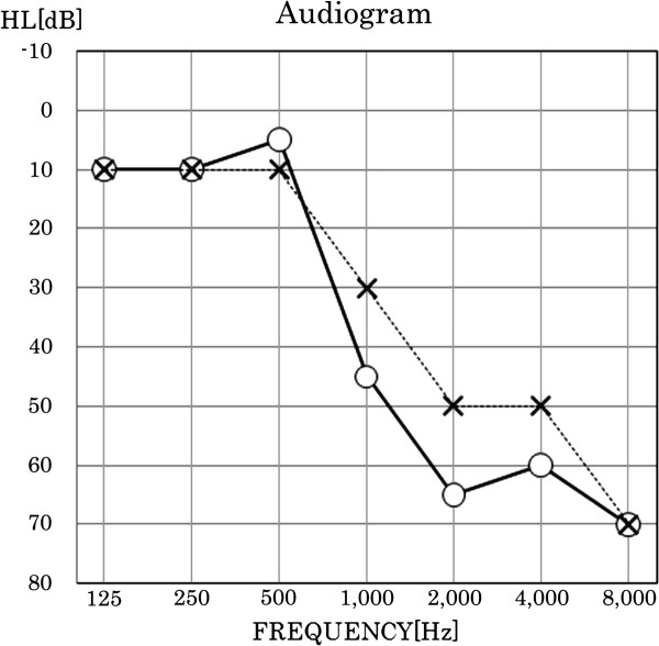
Background: Mutation of the voltage-gated potassium channel KCNQ4 causes DFNA2-type nonsyndromic autosomal dominant sensorineural hearing loss. KCNQ4 is expressed predominantly in the auditory sensory outer hair cells, which are critical for sound amplification.

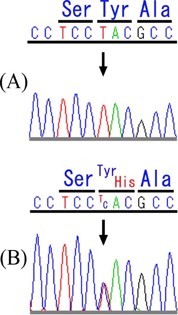
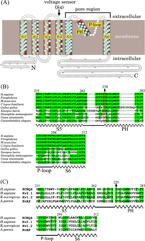
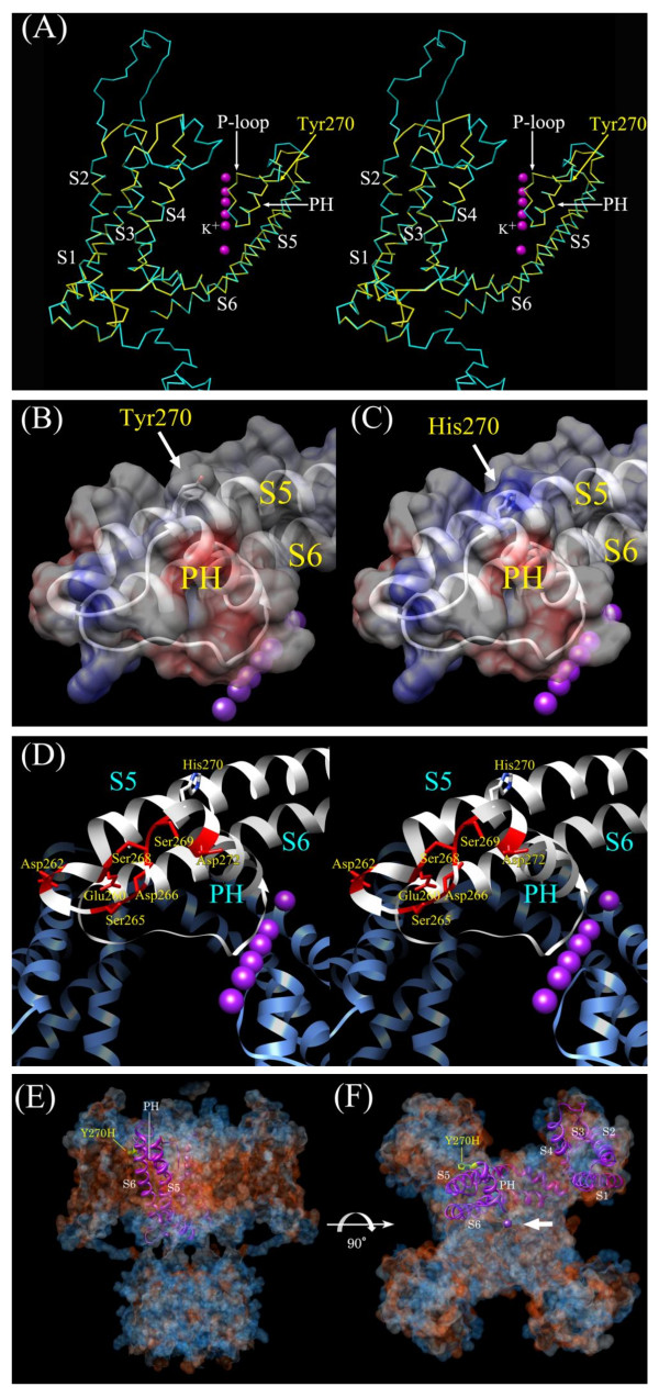
Results: We sequenced KCNQ4 from Japanese patients with sensorineural hearing loss, and identified a novel missense mutation encoding a Tyr270His located at the N-terminus of the highly conserved pore helix sequence. As this patient was not accessible to us and information about them was limited, we used molecular modeling to investigate whether this novel mutation is hypothetically pathogenic. A careful examination of an in silico structural model of the KCNQ4 pore region revealed that the Tyr270His mutation caused an alteration in the electrostatic surface potential of the pore helix.

Conclusion: We propose two possible means by which the Tyr270His mutation causes hearing loss: a positively charged His270 side chain might enhance the helix dipole moment of the pore helix, thereby destabilizing the helix and/or the pore region, or it might disturb transport of K+ through the channel by electrostatic repulsion.


Figures

Figure 1
Figure 1
Partial DNA-sequencing chromatograms of KCNQ4 exon 5 from (A) a subject with normal hearing and (B) the patient with Tyr270His. The position of the heterozygotic T → C mutation at nucleotide position 808 is indicated by the arrow. This mutation was not found in 96 healthy control subjects with normal hearing. No additional mutations were identified in the KCNQ4 coding region of this mutated gene.
Figure 2
Figure 2
An audiogram from the patient with Tyr270His. Open circles connected by the solid line are the hearing levels for the right ear. Crosses connected by the dotted line are the hearing levels for the left ear.
Figure 3
Figure 3
Schematic topology and partial protein sequence of KCNQ4. (A) Schematic topology represents six transmembrane helices (S1–S6) and the K+-selective channel pore region (S5, PH, P-loop, and S6) of KCNQ4. (B) Sequences of the orthologous KCNQ4 pore region are aligned. Positions highlighted in green indicate the amino acid is the same as in human KCNQ4. The position of Tyr270 is indicated by an arrow. The positions of S5, PH, S6 (wavy lines) and the P-loop (straight line) are shown below the sequences. (C) Alignment of the KCNQ4 pore-region sequence with those of Kv channels deposited in PDB. Positions highlighted in green indicate the amino acid is the same as in human KCNQ4. The sequence of human KCNQ4 is indicated at the top of both alignments.
Figure 4
Figure 4
Structural models of KCNQ4 and Tyr270His. (A) Stereoview of the KCNQ4 α-carbon frame model (yellow) superimposed onto that of Kv1.2 (cyan). The position of Tyr270 is indicated with a yellow arrow. The α-helices, the PH, and the P-loop are also identified. (B) Part of the wild-type KCNQ4 model, and (C) the Tyr270His model overlaid with their corresponding electrostatic surface potentials. The side chains of Tyr270 (B) and His270 (C) are indicated by arrows. Negatively and positively charged residues are depicted respectively in red and blue in the electrostatic potential surface representations. (D) Stereoview of a portion of the ribbon model of the Tyr270His pore region. Residues that are negatively charged or that can form a hydrogen bond and surround His270 are identified and shown in red. (E, F) The Tyr270His ribbon model was superimposed onto (E) a horizontal view of the plasma membrane and (F) an extracellular view with the four rotational axes of the Kv1.2 crystal structure shown as hydrophobicity surface. Hydrophobic and hydrophilic residues are depicted respectively in red and blue in the hydrophobicity surface representations. His270 is shown in yellow. (A-F) K+ in the central pore is shown as a purple sphere and indicated by an arrow in (F).

References

    1. Smith RJ, Bale JF Jr, White KR. Sensorineural hearing loss in children. Lancet. 2005;365:879–890. doi: 10.1016/S0140-6736(05)71047-3. - DOI - PubMed
    1. Morton CC, Nance WE. Newborn hearing screening-a silent revolution. N Engl J Med. 2006;354:2151–2164. doi: 10.1056/NEJMra050700. - DOI - PubMed
    1. Kochhar A, Hildebrand MS, Smith RJ. Clinical aspects of hereditary hearing loss. Genet Med. 2007;9:393–408. doi: 10.1097/GIM.0b013e3180980bd0. - DOI - PubMed
    1. Coucke P, Van Camp G, Djoyodiharjo B, Smith SD, Frants RR, Padberg GW, Darby JK, Huizing EH, Cremers CW, Kimberling WJ. Linkage of autosomal dominant hearing loss to the short arm of chromosome 1 in two families. N Engl J Med. 1994;331:425–431. doi: 10.1056/NEJM199408183310702. - DOI - PubMed
    1. Kubisch C, Schroeder BC, Friedrich T, Lütjohann B, El-Amraoui A, Marlin S, Petit C, Jentsch TJ. KCNQ4, a novel potassium channel expressed in sensory outer hair cells, is mutated in dominant deafness. Cell. 1999;96:437–446. doi: 10.1016/S0092-8674(00)80556-5. - DOI - PubMed

Publication types

Substances

Supplementary concepts