

Editorial
doi: 10.1186/1741-7007-10-22.
Ubiquitin ligases and beyond
- PMID: 22420755
- PMCID: PMC3305657
- DOI: 10.1186/1741-7007-10-22
Item in Clipboard
Editorial
Ubiquitin ligases and beyond
BMC Biol.
.
No abstract available
Figures
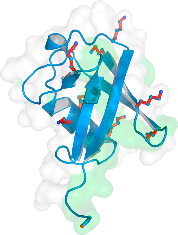
The structure of ubiquitin. Ubiquitin is a small, compact protein characterized by a β-grasp fold. The seven lysines that can be linked to the terminal glycine of another ubiquitin molecule to form poly-ubiquitin chains are colored red. The green shading indicates the hydrophobic patch through which ubiquitin interacts with specific ubiquitin-binding proteins. Image created by Masato Akatsu, Frankfurt University.
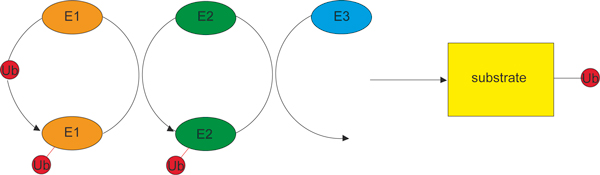
Three enzymes act in sequence to ubiquitinate targets. The E1 enzyme is the activating enzyme, to which ubiquitin is attached in an ATP-dependent reaction by a thioester bond (shown in red). The E2 enzyme is the conjugating enzyme, to which the ubiquitin is transferred from the E1. The E3 is the ubiquitin ligase, which directly or indirectly catalyzes the transfer of the ubiquitin to the target protein (the substrate), with the formation of an isopeptide bond (shown in black).
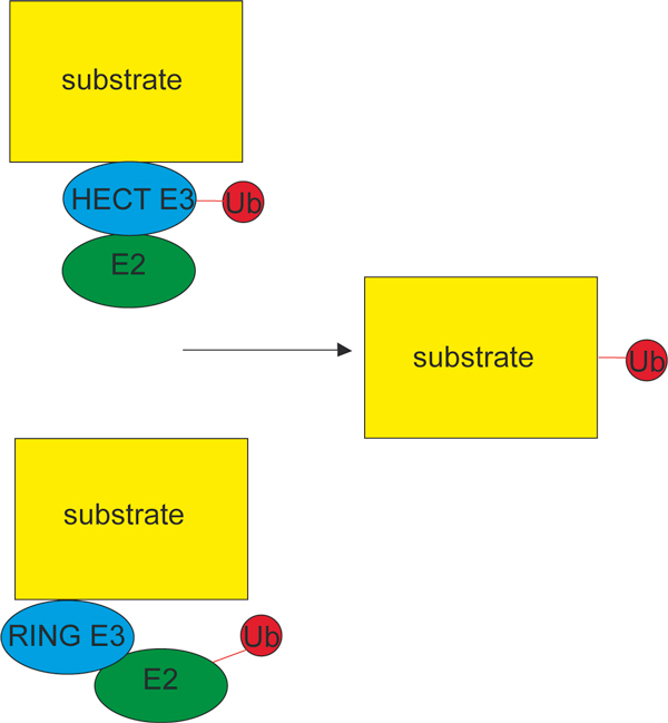
HECT and RING ligases act by different mechanisms. HECT E3 ligases (top) directly catalyze the attachment of ubiquitin to the substrate, whereas in the case of RING ligases (bottom) the ubiquitin is transferred from the E2 which, with the substrate, is bound to the E3. Exactly how the catalytic action of E2 is facilitated by the RING E3 is not known. Reactive thioester bonds are shown in red; the isopeptide bond with the target (substrate) protein is shown in black.
Comment on
-
Cnidocyte discharge is regulated by light and opsin-mediated phototransduction.BMC Biol. 2012 Mar 5;10:17. doi: 10.1186/1741-7007-10-17. BMC Biol. 2012. PMID: 22390726 Free PMC article.
References
Publication types
MeSH terms
Substances
LinkOut - more resources
Full Text Sources
