. 2012 Apr 1;11(7):1364-73.
doi: 10.4161/cc.19765. Epub 2012 Apr 1.

Dual role of hematopoietic progenitor kinase 1 (HPK1) as a positive regulator of 1α,25-dihydroxyvitamin D-induced differentiation and cell cycle arrest of AML cells and as a mediator of vitamin D resistance

Affiliations

Dual role of hematopoietic progenitor kinase 1 (HPK1) as a positive regulator of 1α,25-dihydroxyvitamin D-induced differentiation and cell cycle arrest of AML cells and as a mediator of vitamin D resistance

Xiangwen Chen-Deutsch et al. Cell Cycle. .

Abstract

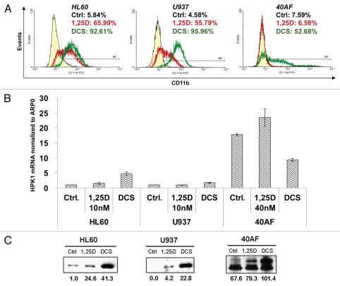
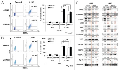
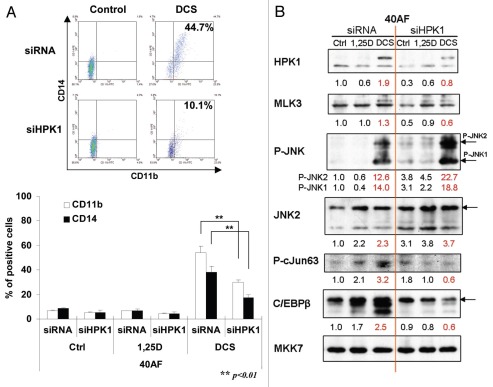
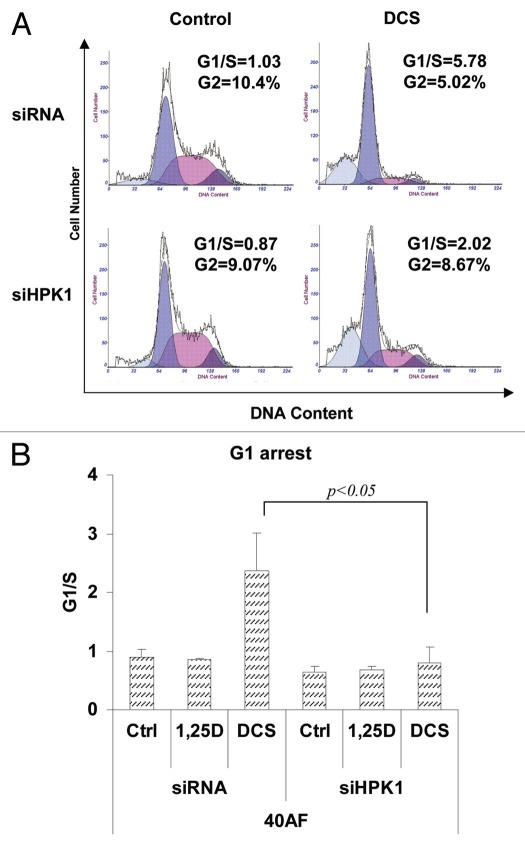
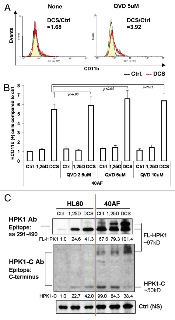
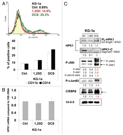
Recent clinical trials aimed at improved treatment of AML by administration of vitamin D derivatives showed unremarkable results, suggesting development of vitamin D resistance in patients' AML blasts. Since mechanisms of vitamin D resistance are not clear, we studied 40AF cells, a subline of HL60 cells that can proliferate in the presence of 1α,25-dihydroxyvitamin D₃ (1,25D). We found that mRNA and protein levels of HPK1, an upstream MAP4 kinase, are dramatically increased in 40AF cells, and HPK1 protein is further increased when the 1,25D resistance of 40AF cells is partially reversed by the addition of carnosic acid and p38MAPK inhibitor SB202190 (DCS cocktail). Knockdown of HPK1 reduces 1,25D/DCS-induced differentiation of both 1,25D-sensitive HL60 and U937 cells and 1,25D-resistant 40AF cells, but the effect of HPK1 knockdown on differentiation-associated G 1 arrest is more apparent in the resistant than the sensitive cells. To explain why 40AF and the intrinsically vitamin D-resistant KG-1a cells can proliferate in the presence of vitamin D, we found that the cleaved HPK1 fragment (HPK1-C) level is high in 40AF and KG-1a cells, but when differentiation is induced by DCS, HPK1-C decreases while full-length (FL)-HPK1 increases. Accordingly, inhibition of proteolysis with the pan-caspase inhibitor Q-VD-OPh reduced HPK1 cleavage and enhanced DCS-induced differentiation of 40AF cells. The results indicate that FL-HPK1 is a positive regulator of vitamin D-induced differentiation in AML cells, but the cleaved HPK1 fragment inhibits differentiation. Thus, high HPK1 cleavage activity contributes to vitamin D resistance, and HPK1 has a dual role in AML cell differentiation.


Figures

Figure 1
Figure 1
Upregulation of HPK1 protein by 1,25D, alone or in combination with enhancers of its action (DCS) parallels the differentiation of AML cells. HL60 (derived from AML-M2) and U937 (derived from AML-M5) represent 1,25D-sensitive cells; 40AF (derived from HL60 maintained in 40 nM 1,25D) represent 1,25D-resistant cells. The cells were treated with 1,25D or DCS (1,25D 10 nM + CA 10 µM + SB 5 µM) for 48 h (sensitive cells) or 72 h (resistant cells). (A) The typical pattern of response to differentiating agents determined by FC for the differentiation marker CD11b. The percentage of positive cells in different treatment groups is indicated in the right top corner of each FC image. (Left peak: control; middle peak: 1,25D; right peak: DCS). Note that in 40AF cells, ‘1,25D’ peak overlaps ‘control peak.’ (B) HPK1 mRNA levels relative to ARP0 as control in different cell lines were determined by real-time RT-PCR. (C) FL-HPK1 protein levels in different cell lines determined by western blots. The OD of each band relative to HL60 untreated (control) cells are displayed below each band. The same amount of total protein extract (40 µg) was loaded for each cell type.
Figure 2
Figure 2
Knockdown of HPK1 in 1,25D-sensitive HL60 and U937 cells decreases 1,25D-induced differentiation, downstream signaling and differentiation-related TFs. Cells were pre-treated with siRNA-HPK1 (siHPK1) or scrambled-siRNA (siRNA) for 24 h, then 10 nM 1,25D was added for another 48 h. Differentiation markers CD11b and CD14 were detected by FC, mean ± SD values are shown, n = 3. Representative images of CD11b (x axis) and CD14 (y axis) determinations are shown in the left side of each figure. (A) HL60 cells; (B) U937 cells. (C) Demonstration of HPK1 decrease, activation of several components of MEKK1-JNK-AP1 signaling cascade and expression of differentiation-related TFs as detected by western blots. Calregulin is a loading control.
Figure 3
Figure 3
HPK1 is required for optimal differentiation of DCS-treated 40AF cells, activates the JNK pathway and leads to increased expression of differentiation-related transcription factors. Cells were pre-treated by siRNA-HPK1 (siHPK1) or scrambled-siRNA (siRNA) for 24 h, 40 nM 1,25D or DCS (1,25D 10 nM + CA 10 µM + SB 5 µM) was added for another 48 h. (A) Expression of CD11b and CD14; Mean ± SD values are shown, n=3. (B) Western blots illustrating HPK1 signaling, with MKK7 serving as loading control, and the effect of HPK1 knockdown on JNK1/2 and the differentiation-related TFs.
Figure 4
Figure 4
Knockdown HPK1 in vitamin D-resistant 40AF cells abrogates the DCS-induced increase in the G1 cell cycle block. Knockdown of HPK1 signaling is described in Figure 3 legend. The cells were stained by propidium iodide and cell cycle distribution was assessed by flow cytometry. (A) Representative images of cell cycle distribution. X-axis: DNA content; y-axis: cell number. The peaks from left to right are: sub-G1, G1, S and G2/M. Note also the abrogation by HPK1 knockdown of DCS-induced G2 compartment depletion. (B) Summary of experiments presented as the G1/S ratio. Mean ± SD values are shown, n = 3.
Figure 5
Figure 5
The pan-caspase inhibitor Q-VD-OPh further enhances DCS-induced differentiation of 40AF cells by inhibition of HPK1 cleavage. (A) Representative images of the changes of CD11b expression in DCS-treated 40AF cells induced by adding 5 µM QVD for 48 h. The ratio of CD11b positive cells between DCS-treated cells and control group is indicated in the right top corner of each image (straight line peak, control; jagged line peak, DCS). (B) Summary of experiments showing the increased expression of CD11b differentiation marker in 1,25D or DCS-treated 40AF cells following addition of increasing concentrations of QVD for 72 h. Mean ± SD values are shown, n = 3. (C) Western blots of HPK1 protein levels in HL60 and 40AF cells treated by 1,25D or DCS for 48 h. HPK1 Ab (epitope: aa 290–490) detects FL-HPK1. HPK1-C Ab (epitope: C-terminus) detects cleaved HPK1-C fragment and FL-HPK1.
Figure 6
Figure 6
DCS treatment of KG-1a cells with innate resistance to 1,25D also reduces the expression of HPK1-C fragments and increases differentiation. (A) CD11b and CD14 expression in KG-1a cells treated by 1 nM 1,25D or DCS (with 1 nM 1,25D) for 72 h. A typical FC image of CD11b (treated for only 48 h) is shown above the summary bar-graph. The left-side peaks overlap and show the negative cell population. The one right-side peak resulting from DCS exposure is apparent. (B) Real-time RT-PCR for HPK1 mRNA in 1,25D or DCS-treated KG-1a cells relative to 18S rRNA as control. There was no significance (p > 0.05) of the mRNA level between control group and 1,25D/DCS group. (C) Western blots of HPK1-JNK-AP-1 signaling pathway in KG-1a cells.

References

    1. Nowak D, Stewart D, Koeffler HP. Differentiation therapy of leukemia: 3 decades of development. Blood. 2009;113:3655–3665. doi: 10.1182/blood-2009-01-198911. - DOI - PMC - PubMed
    1. Miyaura C, Abe E, Kuribayashi T, Tanaka H, Konno K, Nishii Y, et al. 1Alpha,25-Dihydroxyvitamin D3 induces differentiation of human myeloid leukemia cells. Biochem Biophys Res Commun. 1981;102:937–943. doi: 10.1016/0006-291X(81)91628-4. - DOI - PubMed
    1. Koeffler HP. Induction of differentiation of human acute myelogenous leukemia cells: therapeutic implications. Blood. 1983;62:709–721. - PubMed
    1. Huang ME, Ye YC, Chen SR, Chai JR, Lu JX, Zhoa L, et al. Use of all-trans retinoic acid in the treatment of acute promyelocytic leukemia. Blood. 1988;72:567–572. - PubMed
    1. Castaigne S, Chomienne C, Daniel MT, Ballerini P, Berger R, Fenaux P, et al. All-trans retinoic acid as a differentiation therapy for acute promyelocytic leukemia. I. Clinical results. Blood. 1990;76:1704–1709. - PubMed

Publication types

MeSH terms