. 2012 May;31(4):246-52.
doi: 10.1016/j.matbio.2012.02.004. Epub 2012 Mar 6.

Connective tissue mineralization in Abcc6-/- mice, a model for pseudoxanthoma elasticum

Affiliations

Connective tissue mineralization in Abcc6-/- mice, a model for pseudoxanthoma elasticum

N Beril Kavukcuoglu et al. Matrix Biol. 2012 May.

Abstract

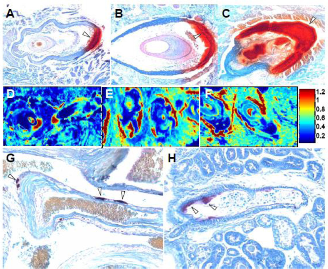
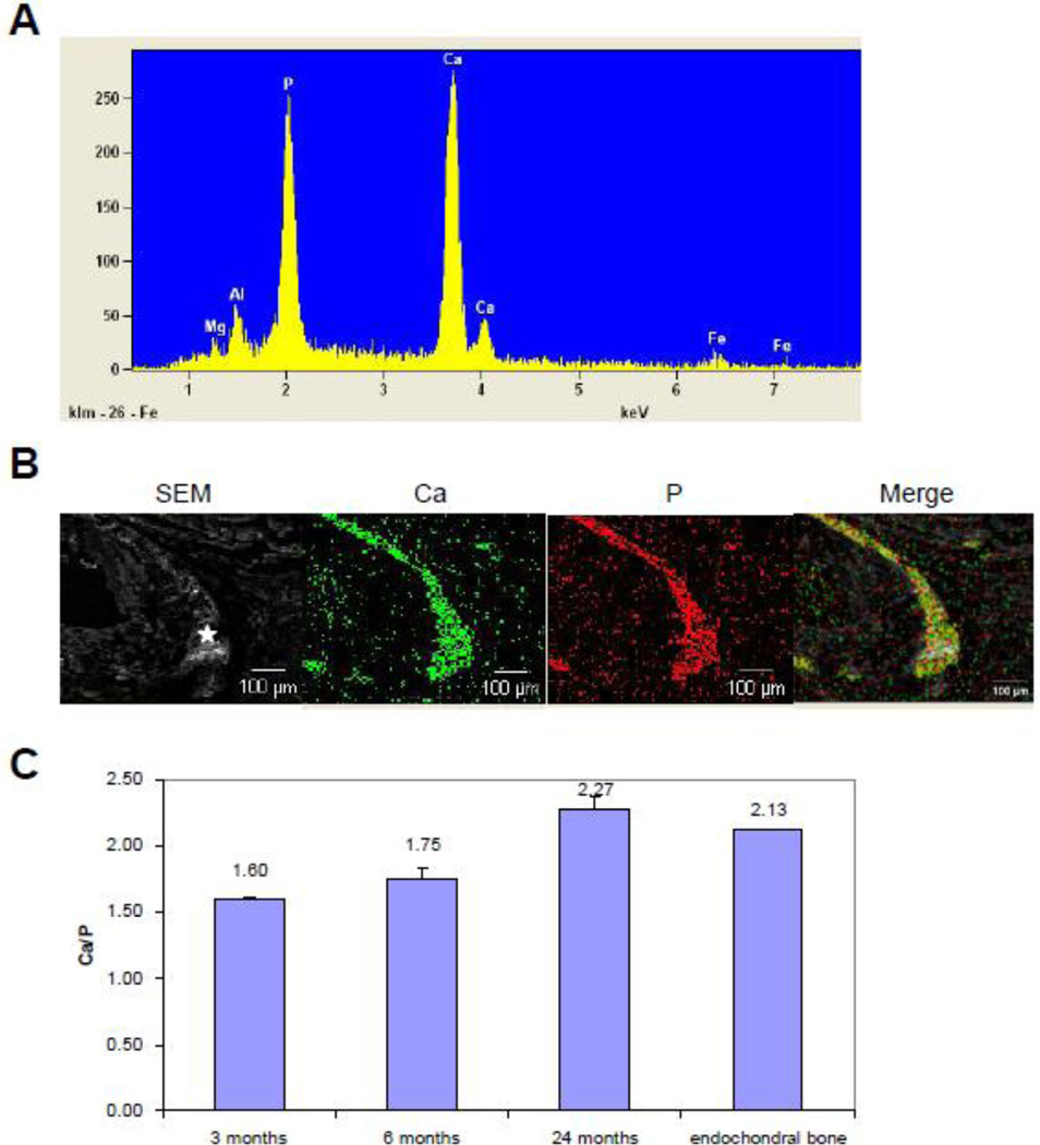
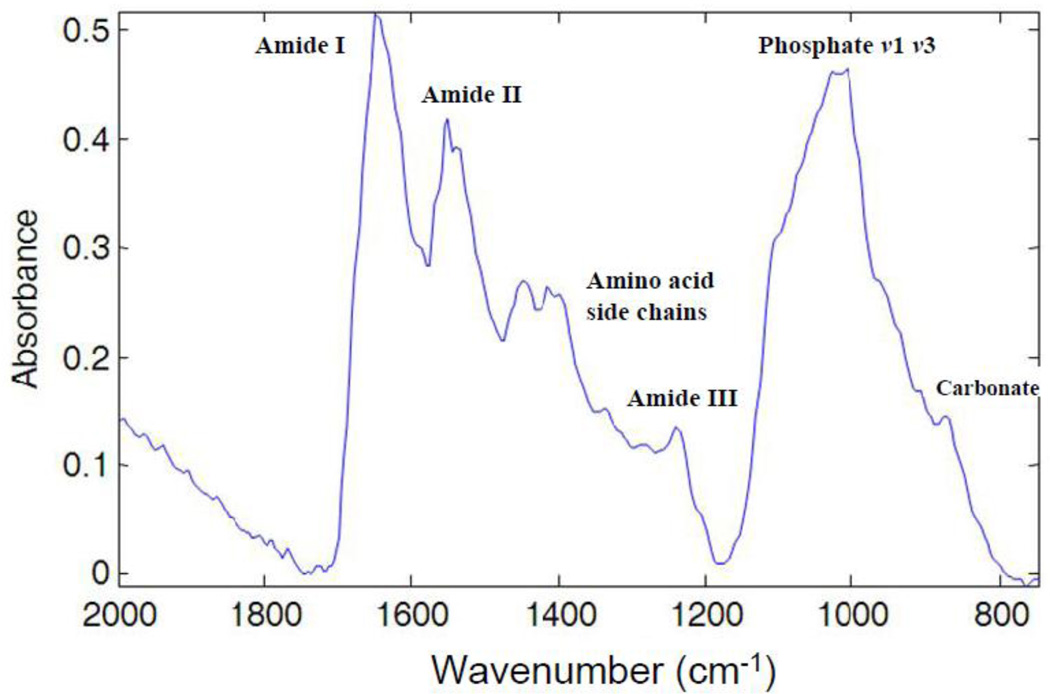
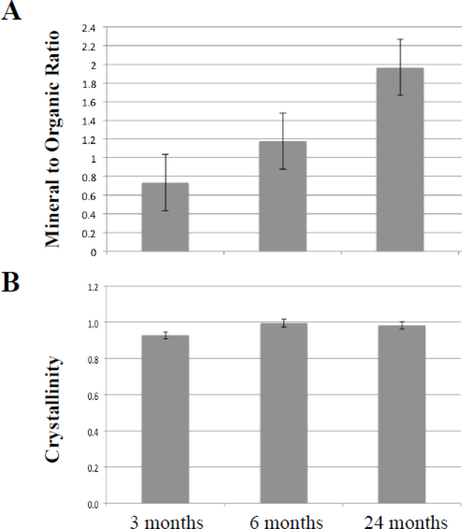
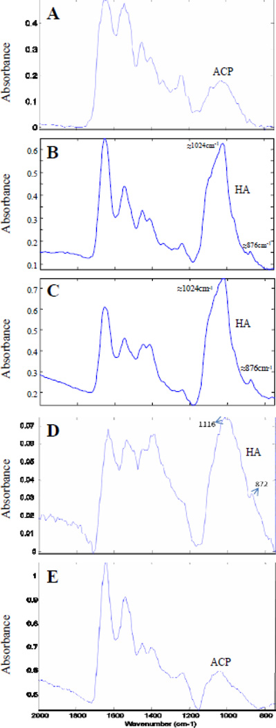
Pseudoxanthoma elasticum (PXE) is a heritable multisystem disorder characterized by ectopic mineralization. However, the structure of the mineral deposits, their interactions with the connective tissue matrix, and the details of the progressive maturation of the mineral crystals are currently unknown. In this study, we examined the mineralization processes in Abcc6(-/-) mice, a model system for PXE, by energy dispersive X-ray and Fourier transform infrared imaging spectroscopy (FT-IRIS). The results indicated that the principal components of the mineral deposits were calcium and phosphate which co-localized within the histologically demonstrable lesions determined by topographic mapping. The Ca/P ratio increased in samples with progressive mineralization reaching the value comparable to that in endochondral bone. A progressive increase in mineralization was also reflected by increased mineral-to-matrix ratio determined by FT-IRIS. Determination of the mineral phases by FT-IRIS suggested progressive maturation of the mineral deposits from amorphous calcium phosphate to hydroxyapatite. These results provide critical information of the mechanisms of mineralization in PXE, with potential pharmacologic implications.


Figures

Figure 1
Figure 1
Ectopic mineralization of the connective tissue capsule surrounding the vibrissae (A–C), and blood vessels in heart (G) and kidney (H), and FT-IRIS images of representative vibrissae samples from mice of different ages, based on the ratio of mineral to matrix peak heights at 1024 and 1660 cm−1. Tissues were isolated from Abcc6−/− mice and processed for staining with Alizarin Red. Note the mineralization was indicated with open arrowheads. Note the progressive mineralization in the connective tissue capsule surrounding the vibrissae in 3 months (A,D), 6 months (B,E), and 24 month old mice (C,F), as well as mineralization in the blood vessels of heart and kidney at 24 months of age (G,H). The color scale represents the ratio of the heights of the absorbance bands of mineral to matrix where red indicates the highest and dark blue indicates the lowest.
Figure 2
Figure 2
Characterization of the mineral deposits by EDAX (A), and topographic mapping (B), and the relative content of calcium and phosphate (C). The results indicated dominant peaks corresponding to calcium and phosphate (A), and these ions co-localize in the mineralized connective tissue capsule (B). The ratio of calcium/phosphate increases from 3 months to 24 months old mice, the latter values approximating those measured in endochondral bone (C).
Figure 3
Figure 3
Illustration of a typical FT-IRIS spectrum of mineralized vibrissae. The bands associated with the vibrations of organic bonds from proteins are shown as amide I (C=O stretch) centered at 1650 cm−1, amide II (C-N stretch and N-H in-plane bending mode) centered at 1550cm−1, amino acid side chain vibrations due to C-H bending motions (overlapping bands 1500–1360 cm−1) and amide III combination band centered around 1250 cm−1, and absorbances from carbonated calcium phosphates as phosphate ν1, ν3 (1200–900 cm−1), and carbonate ν2 (890–840 cm−1).
Figure 4
Figure 4
A. Peak height ratios of the highest absorbance associated with the mineral phase to the absorbance associated with the organic matrix (amide I) in mineral deposits in the connective tissue sheath of vibrissae. The values are mean ± S.E., n = 6–7 per group. B. Peak height ratios of the absorbance at 1030 cm−1 and 1020 cm−1 representing the mineral crystallinity. The values are ± S.E. from 3–9 determinations per group.
Figure 5
Figure 5
Representative FT-IRIS spectra from the mineralized region of tissues from Abcc6−/− mice of varying ages. A. Three months, vibrissae; spectral absorbance typical of amorphous calcium phosphate (ACP). B. Six months, vibrissae; spectral absorbance typical of poorly crystalline hydroxyapatite (HA). C. Twenty four months vibrissae; spectral absorbance typical of poorly crystalline HA. D. Twenty four months, blood vessels in heart; spectral absorbance typical of poorly crystalline HA. E. Twenty four months, blood vessels in kidney; spectral absorbance typical of amorphous calcium phosphate (ACP).

References

    1. Anderson HC, Reynolds JJ. Pyrophosphate stimulation of calcium uptake into cultured embryonic bones. Fine structure of matrix vesicles and their role in calcification. Dev. Biol. 1973;34:211–227. - PubMed
    1. Boskey A, Pleshko Camacho N. FT-IR imaging of native and tissue-engineered bone and cartilage. Biomaterials. 2007;28:2465–2478. - PMC - PubMed
    1. Boskey AL, Mendelsohn R. Infrared spectroscopic characterization of mineralized tissues. Vib. Spectrosc. 2005;38:107–114. - PMC - PubMed
    1. Camacho NP, Carroll P, Raggio CL. Fourier transform infrared imaging spectroscopy (FT-IRIS) of mineralization in bisphosphonate-treated oim/oim mice. Calcif. Tissue Int. 2003;72:604–609. - PubMed
    1. Chua-Anusorn W, Webb J. Infrared spectroscopic studies of nanoscale iron oxide deposits isolated from human thalassemic tissues. J. Inorg. Biochem. 2000;79:303–309. - PubMed

Publication types

MeSH terms

LinkOut - more resources