Review
. 2012 May;19(5):735-42.
doi: 10.1038/cdd.2012.25. Epub 2012 Mar 16.

How apoptotic cells aid in the removal of their own cold dead bodies

Affiliations
Review

How apoptotic cells aid in the removal of their own cold dead bodies

G Wickman et al. Cell Death Differ. 2012 May.

Abstract

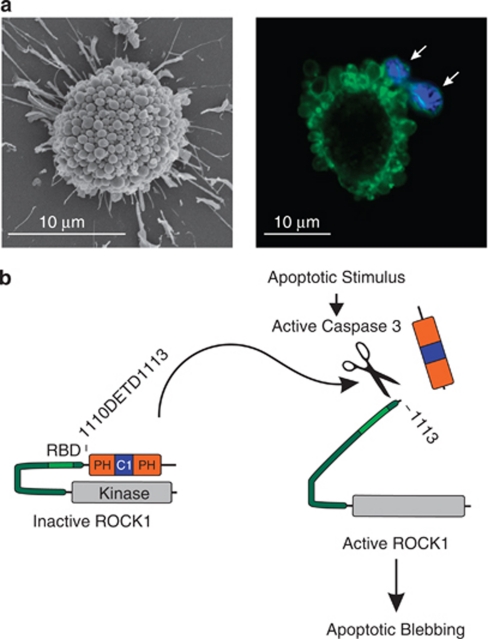
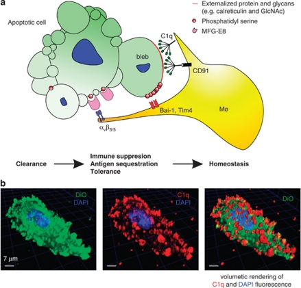
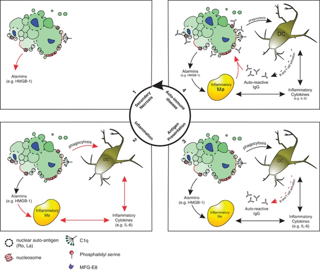
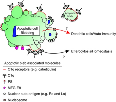
Apoptotic cell clearance facilitates the removal of aged, damaged, infected or dangerous cells although minimizing perturbation of surrounding tissues, and is a vital process in the development and homeostasis of multicellular organisms. Importantly, failure to correctly execute programmed cell death and subsequent corpse clearance is broadly associated with chronic inflammatory and/or autoimmune diseases such as systemic lupus erythematosus. Apoptotic cells develop dramatic morphological changes including contraction, membrane blebbing and apoptotic body formation, which were among the first and most readily identifiable features of cellular suicide. However, understanding the purpose of apoptotic cell morphological changes has proven to be elusive, and recent studies have made somewhat surprising, and occasionally opposing, conclusions about the contribution of blebbing to phagocytic clearance and prevention of inflammatory/autoimmune disease. We review the evidence indicating how apoptotic blebs actively promote corpse recognition, uptake, and generation of auto-reactive antibodies.


Figures

Figure 1
Figure 1
Apoptotic blebbing and ROCK1 cleavage. (a) Representative scanning electron micrograph of apoptotic mouse embryonic fibroblast (MEF) with many surface blebs (left panel). Representative confocal image of apoptotic MEF with multiple blebs containing condensed chromatin (right panel, white arrows). Chromatin is stained with DAPI (blue) and cellular membrane is stained with lipid dye DiO (G=green). (b) Schematic of ROCK cleavage by caspase 3 at D1113. Cleavage yields a constitutively active kinase responsible for apoptotic blebbing. RBD, Rho-binding domain; PH, pleckstrin homology; C1, cysteine rich domain
Figure 2
Figure 2
Blebs focalize factors that promote clearance. (a) Illustration of an apoptotic cell in complex with a macrophage. Apoptotic cell (green) externalizes multiple factors including phosphatidylserine, intracellular proteins and glycan groups. These externalized factors can bind serum proteins C1q and MFG-E8. These membrane modifications enable rapid clearance by acting as ‘Eat-me' signals for surveilling macrophages (Mø,yellow), thus maintaining tissue homeostasis. (b) 3 dimensional confocal reconstruction and volumetric rendering of apoptotic mouse embryonic fibroblast (MEF). Cellular membrane is stained with lipid dye DiO (green), nucleus is stained with DAPI (blue), and cell is stained with human complement C1q (red)
Figure 3
Figure 3
Schematic illustration of failed efferocytosis leading to auto-immune disease. Failure to clear apoptotic cells leads to secondary necrosis and the eventual release of alarmins such as HMGB-1 (panel 1, top left). The secondarily necrotic cell triggers the release of pro-inflammatory cytokines by HMGB-1 stimulation of macrophages (Mø) or following phagocytosis by DCs. (panel 2, bottom-left panel). In the inflammatory context, DCs present ingested apoptotic protein and express co-stimulatory molecules triggering B and T-cell activation leading to the production of auto-reactive antibodies (IgG) (panel 3, bottom right). Auto-reactive antibodies opsonise apoptotic cells and trigger further inflammatory responses characteristic of auto-immune disease (panel 4, top right)
Figure 4
Figure 4
Apoptotic blebs are repositories of membrane modifications. Illustration of apoptotic cell with numerous blebs concentrating ‘Eat-me' signals as well as nuclear auto-antigens. The relative importance of apoptotic blebs for efficient efferocytosis and consequent maintenance of homeostasis versus the presentation of nuclear autoantigens to DCs leading to the development of autoimmunity is unclear, and likely awaits the development of in vivo models deficient in apoptotic blebbing

References

    1. Charras GT, Coughlin M, Mitchison TJ, Mahadevan L. Life and Times of a Cellular Bleb. Biophys J. 2008;94:1836–1853. - PMC - PubMed
    1. Charras GT, Yarrow JC, Horton MA, Mahadevan L, Mitchison TJ. Non-equilibration of hydrostatic pressure in blebbing cells. Nature. 2005;435:365–369. - PMC - PubMed
    1. Charras GT, Hu CK, Coughlin M, Mitchison TJ. Reassembly of contractile actin cortex in cell blebs. J Cell Biol. 2006;175:477–490. - PMC - PubMed
    1. Croft DR, Coleman ML, Li S, Robertson D, Sullivan T, Stewart CL, et al. Actin-myosin-based contraction is responsible for apoptotic nuclear disintegration. J Cell Biol. 2005;168:245–255. - PMC - PubMed
    1. Orlando KA, Stone NL, Pittman RN. Rho kinase regulates fragmentation and phagocytosis of apoptotic cells. Exp Cell Res. 2006;312:5–15. - PubMed

Publication types