. 2012 Oct;19(10):1582-9.
doi: 10.1038/cdd.2012.33. Epub 2012 Mar 16.

Excitotoxic stimulus stabilizes PFKFB3 causing pentose-phosphate pathway to glycolysis switch and neurodegeneration

Affiliations

Excitotoxic stimulus stabilizes PFKFB3 causing pentose-phosphate pathway to glycolysis switch and neurodegeneration

P Rodriguez-Rodriguez et al. Cell Death Differ. 2012 Oct.

Abstract

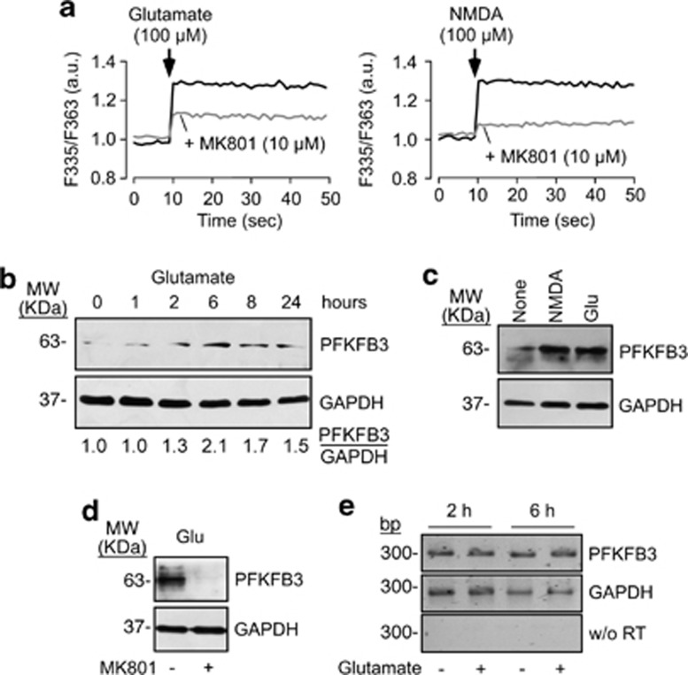
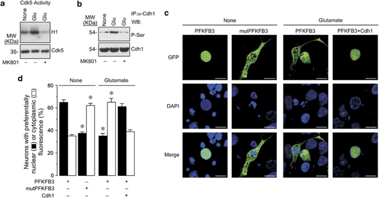
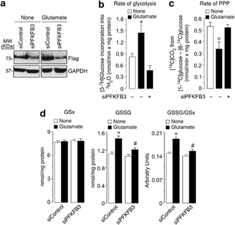
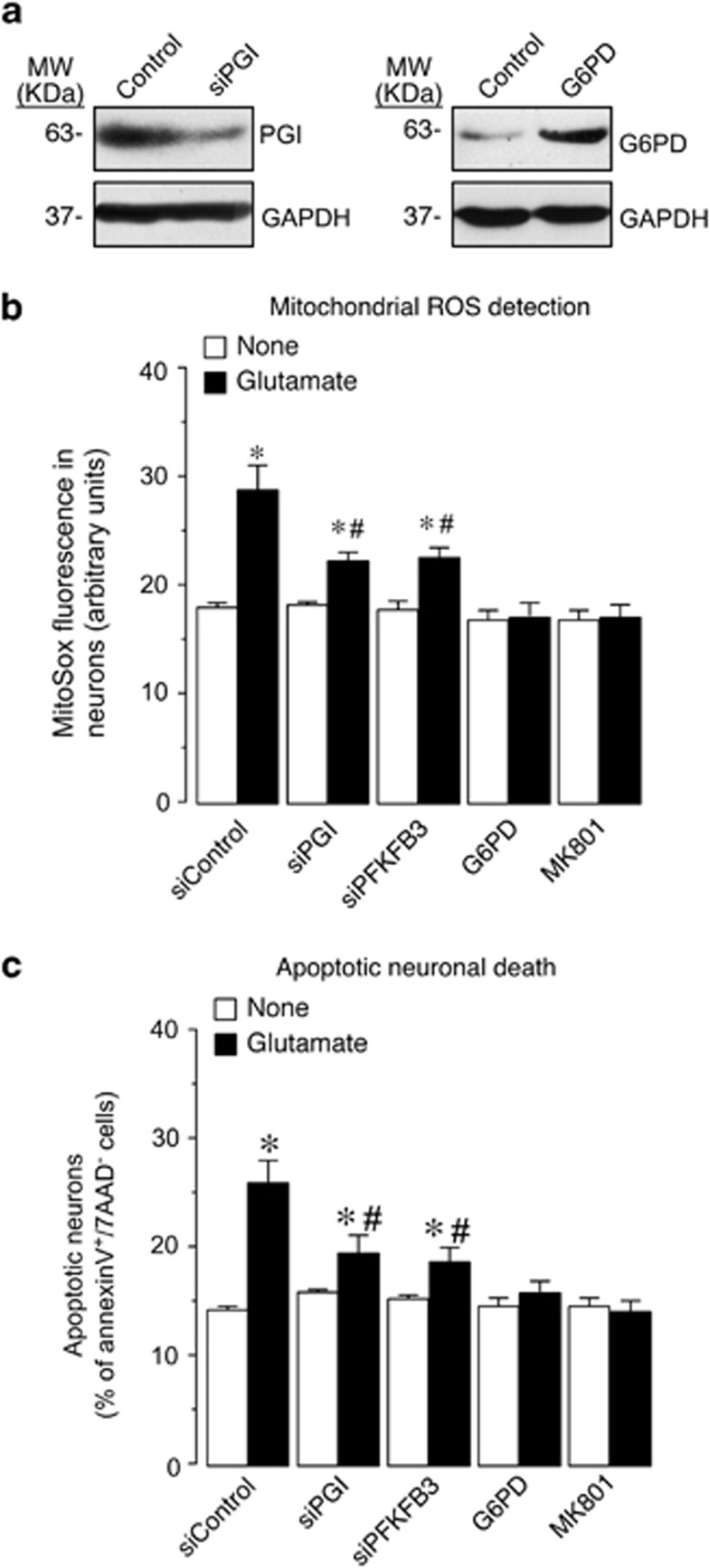
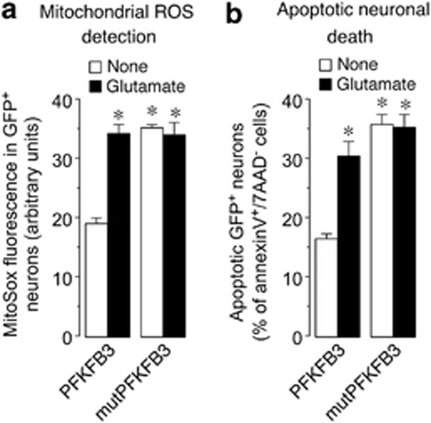
6-Phosphofructo-2-kinase/fructose-2,6-bisphosphatase-3 (PFKFB3) is a master regulator of glycolysis by its ability to synthesize fructose-2,6-bisphosphate, a potent allosteric activator of 6-phosphofructo-1-kinase. Being a substrate of the E3 ubiquitin ligase anaphase-promoting complex-Cdh1 (APC(Cdh1)), PFKFB3 is targeted to proteasomal degradation in neurons. Here, we show that activation of N-methyl-D-aspartate subtype of glutamate receptors (NMDAR) stabilized PFKFB3 protein in cortical neurons. Expressed PFKFB3 was found to be mainly localized in the nucleus, where it is subjected to degradation; however, expression of PFKFB3 lacking the APC(Cdh1)-targeting KEN motif, or following NMDAR stimulation, promoted accumulation of PFKFB3 and its release from the nucleus to the cytosol through an excess Cdh1-inhibitable process. NMDAR-mediated increase in PFKFB3 yielded neurons having a higher glycolysis and lower pentose-phosphate pathway (PPP); this led to oxidative stress and apoptotic neuronal death that was counteracted by overexpressing glucose-6-phosphate dehydrogenase, the rate-limiting enzyme of the PPP. Furthermore, expression of the mutant form of PFKFB3 lacking the KEN motif was sufficient to trigger oxidative stress and apoptotic death of neurons. These results reveal that, by inhibition of APC(Cdh1), glutamate receptors activation stabilizes PFKFB3 thus switching neuronal metabolism leading to oxidative damage and neurodegeneration.


Figures

Figure 1
Figure 1
Activation of NMDAR stabilizes PFKFB3 protein in neurons. (a) Incubation of rat primary cortical neurons with glutamate (left panel) or NMDA (right panel) increased the ratio of Fura-2-dependent fluorescence (at 510 nm) obtained after excitation at 335/363 nm (F335/F363), indicating an increase in intracellular Ca2+. MK801 (10 μM) partially prevented glutamate-induced changes in F335/F363 ratio and most of NMDA-dependent F335/F363 ratio changes. (b) Incubation of neurons with glutamate (100 μM/15 min) triggered time-dependent increase in PFKFB3 protein, which was maximal after 6 h. (c) NMDA (100 μM/15 min) mimicked glutamate at increasing PFKFB3. (d) NMDA receptor antagonist, MK801 (10 μM), prevented glutamate-mediated increase in PFKFB3. (e) Glutamate (100 μM/15 min) did not change PFKFB3 mRNA levels, as revealed by the unaltered intensity of the predicted 300 bp band after reverse-transcription of total RNA samples, followed by polymerase chain reaction (RT-PCR) using specific oligonucleotides for PFKFB3; GAPDH (279 bp band) was used as loading control; the black/white inverted images of the agarose gels are shown; w/o RT, RT-PCR for PFKFB3 without reverse transcriptase
Figure 2
Figure 2
Glutamate-mediated PFKFB3 stabilization occurs via Cdk5-mediated inhibition of APCCdh1 activity. (a) Glutamate treatment (100 μM/15 min) increased, after 1 h, Cdk5-mediated H1 phosphorylation in rat primary cortical neurons; this effect was fully abolished by MK801 (10 μM). (b) Cdh1 is phosphorylated 6 h after glutamate treatment (100 μM/15 min), an effect that was prevented by MK801 (10 μM). (c) Confocal microscopy images of neurons transfected with GFP-PFKFB3 reveals its nuclear localization. Glutamate promotes PFKFB3 accumulation, as revealed by its spread (nuclear plus cytosolic) localization; Cdh1 overexpression prevented this effect. GFP-PFKFB3 mutated on its KEN box (KEN → AAA; mut-PFKFB3) showed the spread-like localization, regardless of glutamate treatment. (d) Percentage of neurons showing nuclear or spread GFP-PFKFB3 localization in the experiments shown in c; these data were obtained by analyzing ∼30 neurons per condition per neuronal preparation (n=4). *P<0.05 versus the corresponding (nuclear or cytoplasmic) PFKFB3-none condition (ANOVA)
Figure 3
Figure 3
Glutamate stimulates PFKFB3-dependent increase in glycolysis, a decrease in PPP and promotes glutathione oxidation in neurons. (a) Incubation of GFP-PFKFB3-expressing neurons with glutamate (100 μM/15 min) induced, 6 h after treatment, PFKFB3 accumulation in control, siRNA-treated neurons (siControl), as revealed by an anti-GFP (Flag) antibody; transfection of neurons with an siPFKFB3 efficiently reduced PFKFB3 protein and prevented glutamate-induced PFKFB3 accumulation. (b) Incubation of neurons with glutamate (100 μM/15 min) increased, after 6 h, the rate of glycolysis, as assessed by the determination of [3-3H]glucose incorporation into 3H2O; this effect was abolished by preventing PFKFB3 accumulation in neurons previously transfected with siPFKFB3. (c) Glutamate treatment decreased, after 6 h, the rate of the PPP, as assessed by the determination of the difference between 14CO2 produced by [1-14C]glucose and that of [6-14C]glucose; this effect was abolished by siPFKFB3. (d) Glutamate treatment did not change GSx (left panel), but it increased GSSG (middle panel) and the oxidized glutathione redox status (GSSG/GSx; right panel); these effects were partially prevented by siPFKFB3. *P<0.05 versus none; #P<0.05 versus the corresponding siControl (ANOVA)
Figure 4
Figure 4
NMDAR activation triggers oxidative stress and apoptotic death by switching PPP to glycolysis. (a) Transfection of neurons with an siRNA against PGI (siPGI), efficiently knocked down PGI protein abundance (left panel). Transfection of neurons with the full-length DNA encoding G6PD efficiently increased G6PD protein abundance (right panel). (b) Glutamate treatment (100 μM/15 min) increased ROS in neurons, as assessed by MitoSox fluorescence by flow cytometry; this effect was prevented by knocking down PGI (siPGI) or PFKFB3 (siPFKFB3), overexpressing G6PD, or blocking NMDAR with MK801 (10 μM). (c) Glutamate treatment increased apoptotic neuronal death, as assessed by annexin V+/7-AAD fluorescence by flow cytometry; this effect was prevented by silencing PGI (siPGI) or PFKFB3 (siPFKFB3), overexpressing G6PD or blocking NMDAR with MK801. *P<0.05 versus none; #P<0.05 versus siControl (glutamate; 5 × 10⁴ events were acquired in triplicate; results mean±S.E.M. from three independent neuronal preparations, n=3; ANOVA)
Figure 5
Figure 5
Expression of APCCdh1-insensitive PFKFB3 mimics glutamate at causing oxidative stress and neuronal death. (a) Glutamate treatment (100 μM/15 min) increased ROS in neurons transfected with low levels of wild-type PFKFB3 cDNA; transfection of neurons with identical cDNA amounts of the KEN box-mut-PFKFB3 increased ROS to similar levels to those triggered by glutamate; glutamate did not further enhance ROS in neurons expressing mut-PFKFB3. (b) Glutamate increased apoptotic death of neurons transfected with low levels of PFKFB3 cDNA; transfection of neurons with identical cDNA amounts of mut-PFKFB3 increased apoptotic death to similar levels to those triggered by glutamate; glutamate did not further enhance apoptotic death in neurons expressing mut-PFKFB3. *P<0.05 versus none (PFKFB3; 3 × 10⁴ events were acquired in triplicate; results mean±S.E.M. from three independent neuronal preparations, n=3; ANOVA)

References

    1. Papadia S, Soriano FX, Leveille F, Martel MA, Dakin KA, Hansen HH, et al. Synaptic NMDA receptor activity boosts intrinsic antioxidant defenses. Nat Neurosci. 2008;11:476–487. - PMC - PubMed
    1. Hardingham GE, Bading H. Synaptic versus extrasynaptic NMDA receptor signalling: implications for neurodegenerative disorders. Nat Rev Neurosci. 2010;11:682–696. - PMC - PubMed
    1. Bossy-Wetzel E, Schwarzenbacher R, Lipton SA. Molecular pathways to neurodegeneration. Nat Med. 2004;10:S2–S9. - PubMed
    1. Bano D, Young KW, Guerin CJ, Lefeuvre R, Rothwell NJ, Naldini L, et al. Cleavage of the plasma membrane Na+/Ca2+ exchanger in excitotoxicity. Cell. 2005;120:275–285. - PubMed
    1. Celsi F, Pizzo P, Brini M, Leo S, Fotino C, Pinton P, et al. Mitochondria, calcium and cell death: a deadly triad in neurodegeneration. Biochim Biophys Acta. 2009;1787:335–344. - PMC - PubMed

Publication types

MeSH terms