Review
. 2013 May;34(5):919-24.
doi: 10.3174/ajnr.A3022. Epub 2012 Mar 15.

MR imaging of papilledema and visual pathways: effects of increased intracranial pressure and pathophysiologic mechanisms

Affiliations
Review

MR imaging of papilledema and visual pathways: effects of increased intracranial pressure and pathophysiologic mechanisms

N Passi et al. AJNR Am J Neuroradiol. 2013 May.

Abstract

Papilledema, defined as swelling of the optic disc, frequently occurs in the setting of increased ICP and in a variety of medical conditions, including pseudotumor cerebri, sinus thrombosis, intracerebral hemorrhage, frontal lobe neoplasms, and Chiari malformation. Noninvasive imaging of the ON is possible by using MR imaging, with a variety of findings occurring in the setting of papilledema, including flattening of the posterior sclera, protrusion of the optic disc, widening of the ONS, and tortuosity of the ON. Early recognition of papilledema and elevated ICP is of paramount importance for ensuring restoration of vision. Newer advanced MR imaging techniques such as fMRI and DTI may prove useful in the future to assess the potential effects of papilledema on retinal and visual pathway integrity.


Figures
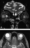
Fig 1.
Fig 1.
ONS enlargement. A, Coronal T2-weighted FSE image demonstrates bilateral widening of the ONS in a 32-year-old woman who presented with acute vision loss and headaches and was found to have papilledema on fundoscopic examination. The ONSs are abnormally increased in size (TR, 6816.7 ms/TE, 84 ms; 3-mm thickness; FOV, 18 cm; matrix, 256 × 224; 4 excitations). B, Axial T2-weighted FSE image demonstrates the widening of the ONS bilaterally in a 31-year-old woman with known papilledema presenting with headaches (TR, 4400 ms/TE, 80 ms; 5-mm thickness; 6-mm spacing; FOV, 24 cm; 320 × 256 matrix).
Fig 2.
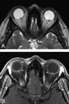
Fig 2.
Optic papilla protrusion. In this T2-weighted axial image, both ON papillae protrude into the vitreous space of the globe. This 32-year-old woman had experienced headaches and vision loss on presentation; she was also found to have papilledema on fundoscopic examination (same patient as in Fig 1A). A, Axial T2-weighted image demonstrates bilateral ON head protrusion (TR, 3000 ms/TE, 84 ms; 5-mm thickness; FOV, 24 cm; matrix, 320 × 256; 1 excitation). B, Corresponding axial T1-weighted postcontrast image demonstrates enhancement of the ON heads (TR, 766.7 ms/TE, 9 ms; 3-mm thickness; FOV, 18 cm; matrix, 320 × 191; 1 excitation).
Fig 3.
Fig 3.
Posterior globe flattening. Flattening of the posterior globe is thought to occur in the same setting as papilledema with increased ICP. In this image, the site of the optic papilla is flattened and the normal globe contour is lost. This 37-year-old woman presented with headache and a sensation of head pressure. She was found to have papilledema and increased CSF opening pressure on lumbar puncture. (TR, 5650 ms/TE, 88 ms; 5-mm thickness; FOV, 24 cm; 320 × 256 matrix).
Fig 4.
Fig 4.
ON tortuosity. Bending of the ON can be seen more prominently in the right ON in this 40-year-old woman with history of headache diagnosed with increased ICP. In this particular case, there is a smear sign, in which orbital fat obscures part of the tortuous ON (TR, 620 ms/TE, 9 ms/TI, 0 ms; 5-mm thickness; 6-mm spacing; FOV, 24 cm; 256 × 192 matrix).

References

    1. Killer HE, Jaggi GP, Miller NR. Papilledema revisited: is its pathophysiology really understood? Clin Experiment Ophthalmol 2009;37:444–47 - PubMed
    1. White LH. Papilledema of ototic origin. Arch Otolaryngol 1925;2:371–78
    1. Holmes G. The prognosis in papilloedema. Br J Ophthalmol 1937;21:337–42 - PMC - PubMed
    1. Hansen HC, Helmke K. Validation of the optic nerve sheath response to changing cerebrospinal fluid pressure: ultrasound findings during intrathecal infusion tests. J Neurosurg 1997;87:34–40 - PubMed
    1. Watanabe A, Kinouchi H, Horikoshi T, et al. . Effect of intracranial pressure on the diameter of the optic nerve sheath. J Neurosurg 2008;109:255–58 - PubMed

MeSH terms