. 2012 Mar 14;483(7389):289-94.
doi: 10.1038/nature10838.

Ancient deuterostome origins of vertebrate brain signalling centres

Affiliations

Ancient deuterostome origins of vertebrate brain signalling centres

Ariel M Pani et al. Nature. .

Abstract

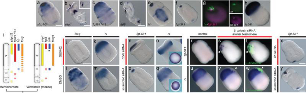
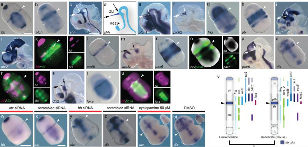
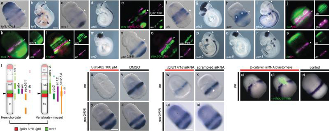
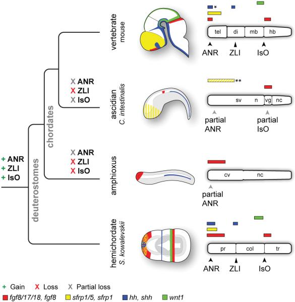
Neuroectodermal signalling centres induce and pattern many novel vertebrate brain structures but are absent, or divergent, in invertebrate chordates. This has led to the idea that signalling-centre genetic programs were first assembled in stem vertebrates and potentially drove morphological innovations of the brain. However, this scenario presumes that extant cephalochordates accurately represent ancestral chordate characters, which has not been tested using close chordate outgroups. Here we report that genetic programs homologous to three vertebrate signalling centres-the anterior neural ridge, zona limitans intrathalamica and isthmic organizer-are present in the hemichordate Saccoglossus kowalevskii. Fgf8/17/18 (a single gene homologous to vertebrate Fgf8, Fgf17 and Fgf18), sfrp1/5, hh and wnt1 are expressed in vertebrate-like arrangements in hemichordate ectoderm, and homologous genetic mechanisms regulate ectodermal patterning in both animals. We propose that these genetic programs were components of an unexpectedly complex, ancient genetic regulatory scaffold for deuterostome body patterning that degenerated in amphioxus and ascidians, but was retained to pattern divergent structures in hemichordates and vertebrates.


Figures

Figure 1
Figure 1. An ANR-like signalling centre in S. kowalevskii
a-h, S. kowalevskii and mouse in situ hybridizations for markers of ANR and telencephalon. S. kowalevskii embryos are at double groove stage (36 hrs), and are shown in dorsal view with anterior to top left, except where noted. Embryos are optically cleared except in o, q insets and r-w. Mouse embryos are at stage ~E8.5. a, S. kowalevskii sfrp1/5 expression. b, frontal view of mouse sfrp1 expression, arrow denotes ANR. Inset shows lateral view. c, S. kowalevskii fgf8/17/18 expression. d, mouse fgf8 expression, arrow denotes ANR. e, S. kowalevskii hh expression. f, S. kowalevskii fgf-Sk1 expression. g, frontal view of double fluorescence in situ hybridization for hh and fgf-Sk1. h, fz5/8 expression. i, AP expression topologies in S. kowalevskii and mouse embryos. Anterior to top. j-k, foxg (j) and rx (k) expression in embryos treated with SU5402. l, m, foxg (l) and rx (m) expression in embryos treated with DMSO. n, expanded apical fgf-Sk1 expression in an embryo injected with fz5/8 siRNA. o, retracted rx expression in an embryo injected with fz5/8 siRNA. p, fgf-Sk1 expression in a siRNA control embryo. q, rx expression in a siRNA control embryo. Insets in o, q show frontal views of uncleared embryos. r, wild type fgf-Sk1 expression. s, fgf-Sk1 expression in descendants of a blastomere injected with β -catenin siRNA. t, merged darkfield/fluorescence images showing clonal descendants of injected cell (green). u, wild-type rx expression. v, rx is not expressed in descendants of a blastomere injected with β -catenin siRNA. w, merged darkfield/fluorescence images showing location of the β-catenin deficient clone (green). x, fgf-Sk1 expression in an embryo injected with hh siRNA. y, fgf-Sk1 expression in a siRNA control embryo. Scale bars = 100 μm in S. kowalevskii, and for mice, 200 μm in (b) and 500 μm in (d). Abbreviations: col, collar; di, diencephalon; hb, hindbrain; mb, midbrain; pr, proboscis; tel, telencephalon; tr, trunk. *, note shh is expressed in the medial ganglionic eminence, near to the ANR.
Figure 2
Figure 2. A ZLI-like signalling centre in S. kowalevskii
a-u, in situ hybridizations for S. kowalevskii and mouse homologs of ZLI and diencephalon markers. Arrowheads mark the proboscis/collar boundary in S. kowalevskii. Mouse images show hemisected heads at E10.5, and dashed lines indicate the ZLI with arrows denoting its extent. a, S. kowalevskii hh expression. b, S. kowalevskii ptch expression (also see Supplementary fig. 2). c, mouse shh expression. d, diagram of shh expression showing ZLI in dark blue. e, mouse ptch1 expression. f, mouse ptch2 expression. g, S. kowalevskii fng expression. h, S. kowalevskii otx expression. i, mouse otx1 expression. j, mouse otx2 expression. k, hh (magenta) and otx (green) are co-expressed at the presumptive proboscis/collar boundary. l, S. kowalevskii wnt8 expression. m, mouse wnt8b expression. n, S. kowalevskii pax6 expression. o, double in situ hybridization showing pax6 expression (fluorescence, green) anterior to hh (colorometric, black). p, mouse pax6 expression. q, S. kowalevskii dlx expression. r, double FISH showing dlx expression (green) in the proboscis base anterior to hh (magenta). s, mouse dlx2 expression. t, S. kowalevskii foxa expression. u, double FISH showing foxa (green) and hh (magenta) expression. v, diagram of AP expression topologies of ZLI and forebrain marker homologs in S. kowalevskii and mice. Six3 expression based on previous data. Anterior to top. w, x, otx siRNA downregulates hh expression at the proboscis/collar boundary (w) relative to a control siRNA (x). y, z, hh siRNA reduces dlx expression in the proboscis base (y) relative to a scrambled siRNA (z). ai, bi, cyclopamine treatment reduces dlx expression in the proboscis base (ai) relative to a control embryo treated with DMSO (bi). Insets show ventral views. Scale bars = 100 μm in S. kowalevskii embryos and 1 mm in mice. Abbreviations: mge, medial ganglionic eminence; pth, prethalamus; th, thalamus; others as described previously.
Figure 3
Figure 3. An IsO-like signalling centre in S. kowalevskii
a-s, in situ hybridizations for S. kowalevskii and mouse homologs of MHB markers. Arrowheads mark the S. kowalevskii collar/trunk coelom boundary the mouse IsO. a, S. kowalevskii fgf8/17/18 expression. b, mouse fgf8 expression. c, S. kowalevskii wnt1 expression. d, mouse wnt1 expression. e, double FISH showing S. kowalevskii wnt1 (green) expressed directly anterior to fgf8/17/18 (magenta). f, S. kowalevskii otx expression. g, mouse otx2 expression. h, S. kowalevskii gbx expression. Asterisks denote endodermal domains. i, mouse gbx2 expression. j, double FISH for S. kowalevskii otx (green) and gbx (magenta). k, double FISH for S. kowalevskii wnt1 (green) and en (magenta). l, double FISH for S. kowalevskii en and otx. m, mouse en2 expression. n, S. kowalevskii pax2/5/8 expression. o, double FISH showing S. kowalevskii pax2/5/8 and en expression. p-r, expression of mouse pax2 (p), pax5 (q), and pax8 (r). s, double FISH for S. kowalevskii th (green) and en (magenta). t, summary of AP expression topologies in hemichordates and mice. Anterior to top. u, en expression is reduced in an embryo treated with SU5402. v, en expression in a DMSO-treated control embryo. w, en expression in an embryo injected with fgf8/17/18 siRNA. x, en expression in an embryo injected with a control siRNA. y, pax2/5/8 expression in an embryo treated with SU5402. z, pax2/5/8 expression in a DMSO-treated control embryo. ai, pax2/5/8 expression in an embryo injected with fgf8/17/18 siRNA. bi, pax2/5/8 expression in an embryo injected with a control siRNA. ci, en is not expressed in descendants of a blastomere injected with β -catenin siRNA. di, merged darkfield/fluorescence images showing location of the β-catenin deficient clone (green). ei, wild-type en expression. Scale bars = 100 μm in S. kowalevskii embryos and 1 mm in mice. Abbreviations as described previously.
Figure 4
Figure 4. Evolutionary gain and loss of ANR, ZLI, and IsO-like genetic programs
Schematic diagrams depicting expression of fgf8, sfrp1, shh, and wnt1 homologs in the mouse brain and ectoderm of C. intestinalis, amphioxus, and S. kowalevskii. Embryos are oriented with anterior to left and dorsal to top. Bar diagrams are oriented with anterior to the left. Diagrams depict only expression domains related to signalling components of vertebrate CNS signalling centres. Abbreviations: cv, cerebral vesicle; n, neck; nc, nerve cord; sv, sensory vesicle; vg, visceral ganglion; others as described previously. Diagrams not to scale. *Note that shh is expressed in the medial ganglionic eminence, adjacent to the ANR.**Sfrp1/5 is expressed in the C. intestinalis anterior ectoderm from 64-cells up to neurulation, but is then downregulated in the anterior ectoderm and CNS (depicted as yellow stripes).

Comment in

References

    1. Echevarria D, Vieira C, Gimeno L, Martinez S. Neuroepithelial secondary organizers and cell fate specification in the developing brain. Brain Res Brain Res Rev. 2003;43:179–191. doi:S0165017303002078 [pii] - PubMed
    1. Wilson SW, Houart C. Early steps in the development of the forebrain. Dev Cell. 2004;6:167–181. doi:S1534580704000279 [pii] - PMC - PubMed
    1. Wurst W, Bally-Cuif L. Neural plate patterning: upstream and downstream of the isthmic organizer. Nat Rev Neurosci. 2001;2:99–108. doi:10.1038/35053516. - PubMed
    1. Wicht H, Lacalli TC. The nervous system of amphioxus: structure, development, and evolutionary significance. Canadian Journal of Zoology. 2005;83:122–150. doi:10.1139/z04-163.
    1. Lacalli TC. Prospective protochordate homologs of vertebrate midbrain and MHB, with some thoughts on MHB origins. International journal of biological sciences. 2006;2:104–109. - PMC - PubMed

Publication types

Substances