

Judging roughness by sight--a 7-Tesla fMRI study on responsivity of the primary somatosensory cortex during observed touch of self and others
- PMID: 22422484
- PMCID: PMC6869993
- DOI: 10.1002/hbm.22031
Judging roughness by sight--a 7-Tesla fMRI study on responsivity of the primary somatosensory cortex during observed touch of self and others
Abstract
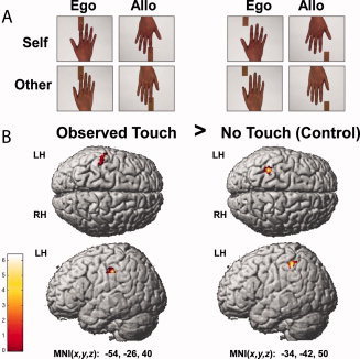
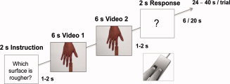
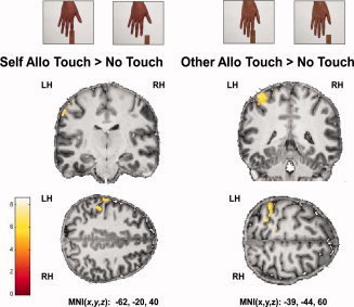
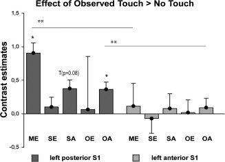
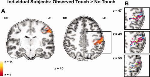
Observing another person being touched activates our own somatosensory system. Whether the primary somatosensory cortex (S1) is also activated during the observation of passive touch, and which subregions of S1 are responsible for self- and other-related observed touch is currently unclear. In our study, we first aimed to clarify whether observing passive touch without any action component can robustly increase activity in S1. Secondly, we investigated whether S1 activity only increases when touch of others is observed, or also when touch of one's own body is observed. We were particularly interested in which subregions of S1 are responsible for either process. We used functional magnetic resonance imaging at 7 Tesla to measure S1 activity changes when participants observed videos of their own or another's hand in either egocentric or allocentric perspective being touched by different pieces of sandpaper. Participants were required to judge the roughness of the different sandpaper surfaces. Our results clearly show that S1 activity does increase in response to observing passive touch, and that activity changes are localized in posterior but not in anterior parts of S1. Importantly, activity increases in S1 were particularly related to observing another person being touched. Self-related observed touch, in contrast, caused no significant activity changes within S1. We therefore assume that posterior but not anterior S1 is part of a system for sharing tactile experiences with others.
Keywords: 7 Tesla; S1; fMRI; observed touch; primary somatosensory cortex; self and other.
Copyright © 2012 Wiley Periodicals, Inc.
Figures
References
-
- Ashburner J, Friston KJ ( 2005): Unified segmentation. Neuroimage 26: 839–851. - PubMed
-
- Avenanti A, Bueti D, Galati G, Aglioti SM ( 2005): Transcranial magnetic stimulation highlights the sensorimotor side of empathy for pain. Nat Neurosci 8: 955–960. - PubMed
-
- Avenanti A, Minio‐Paluello I, Bufalari I, Aglioti SM ( 2006): Stimulus‐driven modulation of motor‐evoked potentials during observation of others' pain. Neuroimage 32: 316–324. - PubMed
-
- Avikainen S, Forss N, Hari R ( 2002): Modulated activation of the human SI and SII cortices during observation of hand actions. Neuroimage 15: 640–646. - PubMed
Publication types
MeSH terms
LinkOut - more resources
Full Text Sources
