. 2012 Nov;56(5):1719-29.
doi: 10.1002/hep.25717. Epub 2012 Aug 27.

Prevention of hepatocellular adenoma and correction of metabolic abnormalities in murine glycogen storage disease type Ia by gene therapy

Affiliations

Prevention of hepatocellular adenoma and correction of metabolic abnormalities in murine glycogen storage disease type Ia by gene therapy

Young Mok Lee et al. Hepatology. 2012 Nov.

Abstract

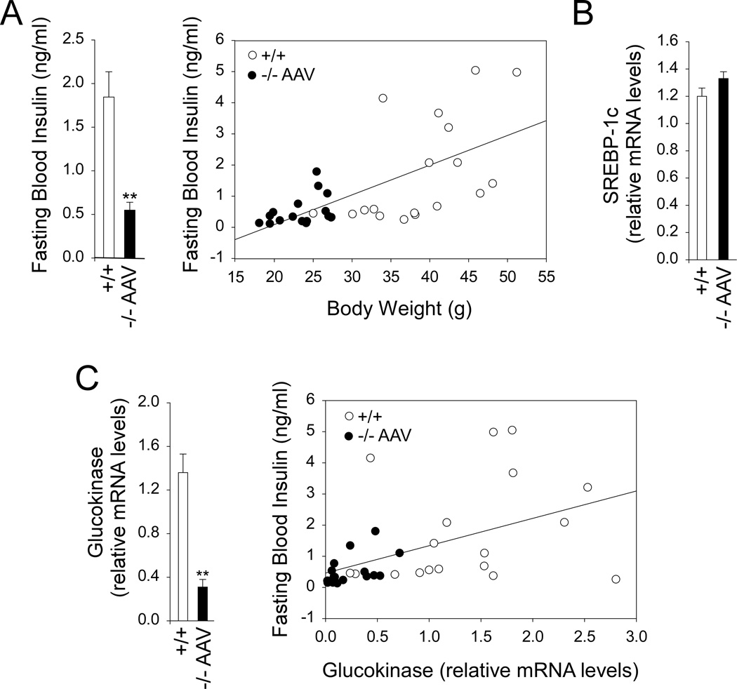
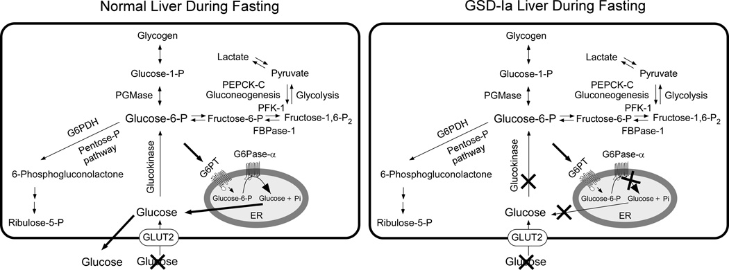
Glycogen storage disease type Ia (GSD-Ia), which is characterized by impaired glucose homeostasis and chronic risk of hepatocellular adenoma (HCA), is caused by deficiencies in the endoplasmic reticulum (ER)-associated glucose-6-phosphatase-α (G6Pase-α or G6PC) that hydrolyzes glucose-6-phosphate (G6P) to glucose. G6Pase-α activity depends on the G6P transporter (G6PT) that translocates G6P from the cytoplasm into the ER lumen. The functional coupling of G6Pase-α and G6PT maintains interprandial glucose homeostasis. We have shown previously that gene therapy mediated by AAV-GPE, an adeno-associated virus (AAV) vector expressing G6Pase-α directed by the human G6PC promoter/enhancer (GPE), completely normalizes hepatic G6Pase-α deficiency in GSD-Ia (G6pc(-/-) ) mice for at least 24 weeks. However, a recent study showed that within 78 weeks of gene deletion, all mice lacking G6Pase-α in the liver develop HCA. We now show that gene therapy mediated by AAV-GPE maintains efficacy for at least 70-90 weeks for mice expressing more than 3% of wild-type hepatic G6Pase-α activity. The treated mice displayed normal hepatic fat storage, had normal blood metabolite and glucose tolerance profiles, had reduced fasting blood insulin levels, maintained normoglycemia over a 24-hour fast, and had no evidence of hepatic abnormalities. After a 24-hour fast, hepatic G6PT messenger RNA levels in G6pc(-/-) mice receiving gene therapy were markedly increased. Because G6PT transport is the rate-limiting step in microsomal G6P metabolism, this may explain why the treated G6pc(-/-) mice could sustain prolonged fasts. The low fasting blood insulin levels and lack of hepatic steatosis may explain the absence of HCA.

Conclusion: These results confirm that AAV-GPE-mediated gene transfer corrects hepatic G6Pase-α deficiency in murine GSD-Ia and prevents chronic HCA formation.


Figures

Fig. 1
Fig. 1
Hepatic G6Pase-α activity and mRNA expression in 70- to 90-week-old wild type and AAV-GPE-treated G6pc−/− mice following a 24-hour fast. Seven 2-week, eleven four-week, one 15-week (♦) and one 30-week (♦♦) old G6pc−/− mice were studied. (A) Hepatic G6Pase-α activity is shown at the indicated ages in weeks (W). The mice are grouped based on their G6Pase-α activity relative to wild type activity as low (AAV-L), medium (AAV-M) and high (AAV-H). (B) Hepatic G6Pase-α mRNA expression and its relationship to G6Pase-α activity in the treated G6pc−/− mice. Data are presented as mean ± SEM. *P < 0.05, **P < 0.005. (C) The relationship between hepatic G6Pase-α mRNA expression and vector genome copy numbers in the treated G6pc−/− mice. (D) Histochemical analysis of hepatic G6Pase-α activity. Freshly sectioned liver specimens were analyzed for G6Pase-α activity using the method of lead trapping of phosphate generated by G6P hydrolysis. Each image represents an individual mouse, so two mice are shown for each treatment. Treatments are indicated by: (−/−), untreated G6pc−/− mice; (+/+), wild type mice; (−/− AAV), G6pc−/− mice infused with various dosages of AAV-GPE. AAV-L (n = 6), AAV-M (n = 9), AAV-H (n = 5) are AAV-GPE-treated G6pc−/− mice expression 3–9%, 22–63%, and 81–128% normal hepatic G6Pase-α activity, respectively.
Fig. 2
Fig. 2
Phenotype analysis of AAV-GPE-treated G6pc−/− mice at age 70 to 90 weeks. (A) Blood glucose, triglyceride, cholesterol, uric acid, and lactic acid levels. (B) Body weight, body length, and BMI. F, females; M, males. (C) Liver weight. Treatments are indicated as: (+/+), wild type mice; (−/− AAV), G6pc−/− mice infused with various dosages of AAV-GPE. AAV-L (n = 6), AAV-M (n = 9), AAV-H (n = 5) are AAV-GPE-treated G6pc−/− mice expressing 3–9%, 22–63%, and 81–128% normal hepatic G6Pase-α activity, respectively. Data are presented as mean ± SEM. *P < 0.05, **P < 0.005.
Fig. 3
Fig. 3
Histological, glycogen, lipid, and COX-2 analyses in the liver of 24-hour fasted wild type and AAV-GPE-infused G6pc−/− mice at age 70 to 90 weeks. Treatments are indicated as: (−/−), untreated G6pc−/− mice; (+/+), wild type mice (n = 20); (−/− AAV) AAV-GPE-treated G6pc−/− mice (n = 20). AAV-L (n = 6), AAV-M (n = 9), AAV-H (n = 5) are the treated G6pc−/− mice expressing 3–9%, 22–63%, and 81–128% normal hepatic G6Pase-α activity, respectively. (A) H&E stained liver sections at original magnifications of ×200 in the treated G6pc−/− mice infused at age 2 or 4 weeks. Each plate represents an individual mouse, so two mice are shown for each treatment. (B) Hepatic glycogen contents. (C) H&E stained liver sections at original magnifications of ×200 in AAV-GPE-treated G6pc−/− mice infused at age 15 or 30 weeks. Duplicated plates are shown for each mouse. (D) Hepatic triglyceride contents. (E) Oil red O staining at original magnifications of ×400. Each plate represents an individual mouse, so two mice are shown for each treatment. (F) Quantification of COX-2 mRNA by real-time RT-PCR. Data are presented as mean ± SEM. *P < 0.05, **P < 0.005.
Fig. 4
Fig. 4
Fasting blood glucose and glucose tolerance profiles. (A) Fasting blood glucose profiles in wild type and AAV-GPE-treated G6pc−/− mice at age 70 to 90 weeks. (B) Fasting blood glucose profiles in untreated G6pc−/− mice at age 6–8 weeks. (C) Glucose tolerance profiles in wild type and the treated G6pc−/− mice at age 70 to 90 weeks. Wild type or the infused G6pc−/− mice were fasted for 6 hours, injected intraperitoneally with 2 mg/g of dextrose, and then sampled for blood every 30 minutes via the tail vein. Data are presented as mean ± SEM. (+/+), wild type mice; (−/−), untreated G6pc−/− mice. AAV-L (n = 6), AAV-M (n = 9), AAV-H (n = 5) are AAV-GPE-treated G6pc−/− mice expression 3–9%, 22–63%, and 81–128% normal hepatic G6Pase-α activity, respectively.
Fig. 5
Fig. 5
Blood insulin and hepatic mRNA levels for SREBP-1c and glucokinase in 70 to 90 week-old wild type and AAV-GPE-treated G6pc−/− mice after 24 hours of fast. (A) Fasting blood insulin levels and its relationship to body weights of the animals. (B) Quantification of SREBP-1c mRNA by real-time RT-PCR. (C) Quantification of glucokinase mRNA and the relationship of fasting blood insulin to hepatic glucokinase mRNA levels. (+/+, ○), wild type mice (n = 20); (−/− AAV, ●) AAV-GPE-treated G6pc−/− mice (n = 20). Data are presented as mean ± SEM. **P< 0.005.
Fig. 6
Fig. 6
Glucose homeostasis and analysis of anti-G6Pase-α antibody in wild type and AAV-GPE-treated G6pc−/− mice. Hepatic levels of free glucose, G6P, mRNA for PEPCK-C, FBPase-1, PGMase, G6PT, PFK-1, and G6PDH were determined in the liver of wild type and the treated G6pc−/− mice after a 24-hour fast at age 70 to 90 weeks. (A) Hepatic free glucose and G6P levels. (B) (C) Quantification of PEPCK-C, FBPase-1, PGMase, G6PT (B), PFK-1, and G6PDH (C) mRNA levels by real-time RT-PCR. (D) Microsomal G6P uptake activity. Data are presented as mean ± SEM. *P< 0.05, **P< 0.005. (E) Analysis of anti-G6Pase-α antibody in the sera of 70- to 90-week-old wild type and AAV-GPE-treated G6pc−/− mice. Microsomal proteins from Ad-human G6Pase-α infected COS-1 cells were electrophoresed through a single 12% polyacrylamide-SDS gel and transferred onto a PVDF membrane. Membrane strips, representing individual lanes on the gel were individually incubated with the appropriate serum. A monoclonal antibody against human G6Pase-α that also recognizes murine G6Pase-α was used as a positive control (lane 1). Treatments are indicated as: (+/+), wild type mice; (−/− AAV), G6pc−/− mice infused with various dosages of AAV-GPE. AAV-L (n = 6), AAV-M (n = 9), AAV-H (n = 5) are the treated G6pc−/− mice expressing 3–9%, 22–63%, and 81–128% normal hepatic G6Pase-α activity, respectively.
Fig. 7
Fig. 7
Pathways for G6P metabolism in normal and GSD-Ia liver during fasting. During fasting, G6P, the end product of gluconeogenesis and glycogenolysis, is transported from the cytoplasm into the lumen of the ER by G6PT. Inside the ER, G6P is hydrolyzed by G6Pase-α and the resulting glucose transported back into the cytoplasm then released into the circulation. In the GSD-Ia liver, which lacks a functional G6Pase-α, ER-localized G6P cannot be converted to glucose, leading to hypoglycemia following a short fast. The GLUT2 transporter, responsible for the transport of glucose in and out of the cell, is shown embedded in the plasma membrane. The G6PT, responsible for the transport of G6P into the ER, and G6Pase-α, responsible for hydrolyzing G6P to glucose and phosphate, are shown embedded in the ER membrane.

Comment in

Similar articles

Cited by

References

    1. Chou JY, Jun HS, Mansfield BC. Glycogen storage disease type I and G6Pase-β deficiency: etiology and therapy. Nat Rev Endocrinol. 2010;6:676–688. - PMC - PubMed
    1. Greene HL, Slonim AE, O'Neill JA, Jr, Burr IM. Continuous nocturnal intragastric feeding for management of type 1 glycogen-storage disease. N Engl J Med. 1976;294:423–425. - PubMed
    1. Chen YT, Cornblath M, Sidbury JB. Cornstarch therapy in type I glycogen storage disease. N Engl J Med. 1984;310:171–175. - PubMed
    1. Labrune P, Trioche P, Duvaltier I, Chevalier P, Odievre M. Hepatocellular adenomas in glycogen storage disease type I and III: a series of 43 patients and review of the literature. J Pediatr Gastroenterol Nutr. 1997;24:276–279. - PubMed
    1. Rake JP, Visser G, Labrune P, Leonard JV, Ullrich K, Smit GP. Glycogen storage disease type I: diagnosis, management, clinical course and outcome. Results of the European Study on Glycogen Storage Disease Type I (ESGSD I) Eur J Pediatr. 2002;161(Suppl 1):S20–S34. - PubMed

Publication types

MeSH terms