. 2012 Jun 15;21(12):2745-58.
doi: 10.1093/hmg/dds102. Epub 2012 Mar 14.

The zinc finger protein ZPR1 is a potential modifier of spinal muscular atrophy

Affiliations

The zinc finger protein ZPR1 is a potential modifier of spinal muscular atrophy

Saif Ahmad et al. Hum Mol Genet. .

Abstract

Spinal muscular atrophy (SMA) is caused by mutation of the Survival Motor Neurons 1 (SMN1) gene and is characterized by degeneration of spinal motor neurons. The severity of SMA is primarily influenced by the copy number of the SMN2 gene. Additional modifier genes that lie outside the SMA locus exist and one gene that could modify SMA is the Zinc Finger Protein (ZPR1) gene. To test the significance of ZPR1 downregulation in SMA, we examined the effect of reduced ZPR1 expression in mice with mild and severe SMA. We report that the reduced ZPR1 expression causes increase in the loss of motor neurons, hypermyelination in phrenic nerves, increase in respiratory distress and disease severity and reduces the lifespan of SMA mice. The deficiency of SMN-containing sub-nuclear bodies correlates with the severity of SMA. ZPR1 is required for the accumulation of SMN in sub-nuclear bodies. Further, we report that ZPR1 overexpression increases levels of SMN and promotes accumulation of SMN in sub-nuclear bodies in SMA patient fibroblasts. ZPR1 stimulates neurite growth and rescues axonal growth defects in SMN-deficient spinal cord neurons from SMA mice. These data suggest that the severity of disease correlates negatively with ZPR1 levels and ZPR1 may be a protective modifier of SMA.


Figures

Figure 1.
Figure 1.
Reduced expression of ZPR1 causes increase in the loss of spinal motor neurons and the severity of disease in mice with mild SMA. (A) Reduced dosage of Zpr1 or Smn genes results in decreased levels of SMN and ZPR1 proteins in the spinal cords. The amounts of ZPR1, SMN and tubulin in spinal cords from 12-month-old mice were examined by immunoblot analysis. The relative change in amounts of proteins was calculated using the amount of each protein normalized to tubulin in wild-type (WT) as reference point (100%). (B) SMA disease carriers and patients (SMA type-I) express low levels of ZPR1. The amounts of SMN, ZPR1 and actin were examined by immunoblot analysis of cell lysates prepared from fibroblasts of non-SMA human (SMN1+/+) (WI-38), carriers of SMA (SMN1/+) mother (GM03814) and father (GM03815) of SMA child (GM03813) and SMA patients (SMN1/−) [GM03813, GM09677 and GM0232A]. The relative amounts of SMN and ZPR1 proteins normalized to β-actin are presented as (mean ± SD; n = 3). (C) ZPR1 deficiency increases defects in the gait of mice with mild SMA (Smn/+). Footprint patterns of 12-month-old WT, Smn/+, Zpr1/+ and [Smn/+; Zpr1/+] littermates (red, fore-paws; blue, hind-paws) were examined. Quantitative analysis of stride length and paw abduction for males (M) and females (F) was performed three times (five mice/group) and results are presented as (mean ± SD). (D) Histochemical staining with H&E and immunohistochemical staining with SMN antibody of the lumbar region of spinal cords from 12-month-old mice (scale bar, 50 µm). (E) Motor neurons were counted in sections of the lumbar (L1–L5) region of spinal cords (mean ± SD; five mice/group) from 12-month-old mice. The loss of motor neurons was calculated using the number of motor neurons in wild-type mice as the reference point (100%).
Figure 2.
Figure 2.
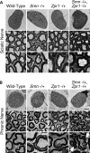
Low levels of ZPR1 cause myelin defects in the PNS of mice with mild SMA. (A) Transverse ultra-thin sections of sciatic nerves of 12-month-old wild-type, Smn/+, Zpr1/+ and [Smn/+; Zpr1/+] mice were examined by TEM. Scale bars are 60 µm (upper), 4 µm (middle) and 2 µm (lower) panels. (B) Transverse ultra-thin sections of phrenic nerves of 12-month-old mice were examined by TEM. Arrows indicate hypermyelination and myelin folding (tomaculi). Scale bars are 60 µm (upper), 4 µm (middle) and 2 µm (lower) panels.
Figure 3.
Figure 3.
Reduced Zpr1 gene dosage increases the severity of disease and decreases the lifespan of mice with severe SMA. (A) ZPR1 deficiency increases weakness and severity in SMA mice. A photograph of 10-day-old littermates, non-SMA (WT) [Smn+/+; SMN2+/+; SMNΔ7+/+; Zpr1+/+], SMA [Smn/−; SMN2+/+; SMNΔ7+/+; Zpr1+/+] and SMA mouse with Zpr1 mutation (SZ) [Smn/−; SMN2+/+; SMNΔ7+/+; Zpr1/+]. Genotypes were examined by PCR. The Quick-Time movie (Supplementary Material) of 10-day-old littermates with WT, SMA and SZ genotypes shows motor activity. (B) Reduced Zpr1 gene dosage causes decreased expression of ZPR1 and SMN in the spinal cord of SMA mice. The amounts of ZPR1, SMN and tubulin in tissue were examined by immunoblot analysis. The change in amounts of proteins was calculated by normalizing to the levels of tubulin in wild-type as the reference point (100%). (C) Reduced expression of ZPR1 increases the severity of disease in mice with SMA. Tail hanging of WT, SMA and SZ mice shows opening (arrowheads) and clasping (arrows) of front and hind limbs, respectively. (D) ZPR1 deficiency causes defects in the growth of mice with SMA. Analysis of growth as total body weights (g) with age (days) in WT (blue triangles), SMA (green squares) and SZ (red circles) mice is shown as growth curves. The results are presented as (mean ± SD, 10 mice/group). (E) Reduced expression of ZPR1 results in the decrease of lifespan in mice with SMA. Kaplan–Meier survival analysis of WT (blue, triangles), SMA mice (green, squares) and SMA mice with Zpr1 mutation (SZ) (red, circles). (F) ZPR1 deficiency causes increase in the loss of spinal cord motor neurons. Histochemical staining with H&E and immunohistochemical staining with SMN antibody of the spinal cords sections from 10-day-old WT, SMA and SZ littermates (scale bar, 50 µm). Bar graph shows the loss of motor neurons in the thoracic region (T9–T12) of spinal cords from SMA and SZ mice compared with WT mice as the reference point (100%) (mean ± SD; five mice/group). (G) The ultra-thin sections of the spinal cords of WT, SMA and SZ littermates were examined by TEM. The arrows indicate heterochromatin condensation in the nucleus. Asterisks indicate void spaces. Scale bar is 2.0 µm.
Figure 4.
Figure 4.
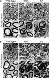
ZPR1 deficiency causes axonal degeneration and hypermyelination in the phrenic nerve of SMA mice. (A) Transverse ultra-thin sections of sciatic nerves of wild-type (WT), mice with SMA (SMA) and SMA mice with Zpr1/+ mutation (SZ) littermates were examined by TEM. The arrows indicate myelin degeneration. Scale bar is 2 µm (upper), 1 µm (lower). (B) Transverse ultra-thin sections of phrenic nerves of WT, SMA and SZ littermates were examined by TEM. Arrowheads indicate hypermyelination and myelin folding (tomaculi). Scale bar is 2 µm (upper), 1 µm (lower).
Figure 5.
Figure 5.
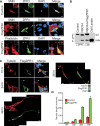
ZPR1 promotes nuclear accumulation of SMN and increases levels of SMN in SMA patient cells. (A) The fibroblasts from non-SMA human (WI-38) (normal) and SMA type-I patient (GM03813) were cultured, mock-transfected (control) and transfected with pcDNA3/FlagZPR1 (Flag-ZPR1), processed and examined by immunofluorescence analysis. The sub-cellular distribution of ZPR1, SMN and FlagZPR1 was examined using monoclonal antibodies to ZPR1, SMN and FLAG (M2) for the detection of recombinant Flag-ZPR1. The colocalization (yellow) and accumulation of ZPR1 (red) and SMN (green) in the nucleus is indicated by arrows. DNA was stained with DAPI (blue). Scale bar is 10 μm. (B) The fibroblasts from normal (WI-38), SMA carriers (GM03814 and GM03815) and SMA patients (GM03813, GM09677 and GM0232A) were infected with empty Ad5CMV (control) and with recombinant adenovirus (Ad5CMV) expressing FlagZPR1 (Flag-ZPR1). The amounts of ZPR1, Flag-ZPR1, SMN, Gemin2 and β-actin in cell lysates were examined by immunoblot analysis. The change in the amounts of proteins (mean ± SD; n = 3) was calculated using the amount of each protein normalized to actin in WI-38 as the reference point (100%).
Figure 6.
Figure 6.
ZPR1 stimulates neuron differentiation and axonal growth in motor neuron-like cells. (A) Colocalization of endogenous and overexpressed ZPR1 with SMN in motor neuron-like (NSC-34) cells. Undifferentiated NSC-34 cells were mock-transfected or transfected with pCDNA3/FlagZPR1. After 24h of transfection, cells were differentiated into neurons. Cells were harvested at 24h after differentiation, fixed and stained with antibodies to SMN (red) and ZPR1-FITC (green) (control), SMN (red) and FLAG (M2)-FITC (green) (center panel), phalloidin (red) and FLAG epitope (M2) (green) (bottom panel). Arrows indicate sub-nuclear bodies. Arrowheads show growth cones. Nuclei were stained with DAPI (blue). Scale bar is 20 μm. (B) Interaction of ZPR1 with SMN in NSC-34 cells. Endogenous and overexpressed ZPR1 proteins were immunoprecipitated with antibodies to ZPR1 and FLAG M2, respectively. Immune complexes were separated by SDS–PAGE and SMN was detected by immunoblot analysis. (C) ZPR1 stimulates axon growth and differentiation in cultured motor neuron-like cells. NSC-34 cells transfected with FlagZPR1 were differentiated into neurons after 24h. Cells were harvested at 12, 24, 36 and 48h post-differentiation, examined by immunofluorescence using antibodies to FLAG (M2) (green) and neuron-specific class-III β-tubulin (red). DNA was stained with DAPI (blue). Arrows indicate nuclear bodies. Arrowheads indicate growth cones. Asterisks indicate untransfected cells. Scale bar is 20 μm. (D) The bar graph shows the average neurite length (mean ± SD, n = 50) in untransfected (control) and transfected (FlagZPR1) neurons.
Figure 7.
Figure 7.
ZPR1 promotes accumulation of SMN in the nucleus and rescues axon length phenotype in SMN-deficient neurons derived from SMA mice. (A) Interaction of ZPR1 with SMN was disrupted in SMA mice. ZPR1 was immunoprecipitated with antibodies to ZPR1 from the brain and spinal cords of normal and SMA mice. Immune complexes were separated by SDS–PAGE, and SMN was detected by immunoblot analysis. (B) Colocalization of endogenous and overexpressed ZPR1 with SMN in spinal cord neurons from non-SMA mice (normal) and SMA mice (SMA) expressing recombinant ZPR1 (SMA+FlagZPR1). Spinal cord explant neuronal cultures were infected with adenovirus, empty virus (Ad5CMV) and with recombinant ZPR1 (Ad5CMV-FlagZPR1), harvested after 48h of infection and stained with antibodies to SMN (red), ZPR1 (green) and FLAG (M2) to detect recombinant Flag-ZPR1 (green). Nuclei were stained with DAPI (blue). Arrows indicate colocalization of ZPR1 and SMN in sub-nuclear bodies. Scale bar is 20 μm. (C) ZPR1 stimulates axonogenesis and rescues the axon length in SMN-deficient spinal motor neurons from SMA mice. Spinal cords neurons from 7-day-old non-SMA (normal) and SMA mice were infected with Ad5CMV-FlagZPR1, fixed after 48h post-infection and stained with antibodies to tubulin (red) and FLAG (M2) to detect recombinant FlagZPR1 (green). Nuclei were stained with DAPI (blue). Asterisks indicate neurons that do not express FlagZPR1. Neurons that express FlagZPR1 are stained in green. Arrows indicate bending and retraction of SMN-deficient spinal cord neurons. Scale bar is 20 μm. (D) The bar graph shows average neurite length [mean ± SEM, n = 30 (cells), 3 mice/group] in untransfected (control) and transfected (FlagZPR1) spinal cord neurons from non-SMA (normal) and SMA mice (SMA).

References

    1. Munsat T.L., Davies K.E. International SMA consortium meeting (26–28 June 1992, Bonn, Germany) Neuromuscul. Disord. 1992;2:423–428. - PubMed
    1. Lefebvre S., Burglen L., Reboullet S., Clermont O., Burlet P., Viollet L., Benichou B., Cruaud C., Millasseau P., Zeviani M., et al. Identification and characterization of a spinal muscular atrophy- determining gene. Cell. 1995;80:155–165. - PubMed
    1. Lefebvre S., Burlet P., Liu Q., Bertrandy S., Clermont O., Munnich A., Dreyfuss G., Melki J. Correlation between severity and SMN protein level in spinal muscular atrophy. Nat. Genet. 1997;16:265–269. - PubMed
    1. Lorson C.L., Hahnen E., Androphy E.J., Wirth B. A single nucleotide in the SMN gene regulates splicing and is responsible for spinal muscular atrophy. Proc. Natl Acad. Sci. USA. 1999;96:6307–6311. - PMC - PubMed
    1. Monani U.R., Lorson C.L., Parsons D.W., Prior T.W., Androphy E.J., Burghes A.H., McPherson J.D. A single nucleotide difference that alters splicing patterns distinguishes the SMA gene SMN1 from the copy gene SMN2. Hum. Mol. Genet. 1999;8:1177–1183. - PubMed

Publication types

MeSH terms

Substances