. 2012 Jun 15;21(12):2759-67.
doi: 10.1093/hmg/dds104. Epub 2012 Mar 14.

Absence of triadin, a protein of the calcium release complex, is responsible for cardiac arrhythmia with sudden death in human

Affiliations

Absence of triadin, a protein of the calcium release complex, is responsible for cardiac arrhythmia with sudden death in human

Nathalie Roux-Buisson et al. Hum Mol Genet. .

Abstract

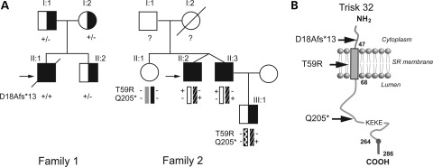
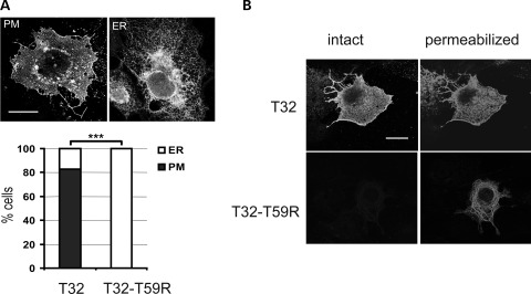
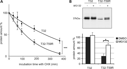
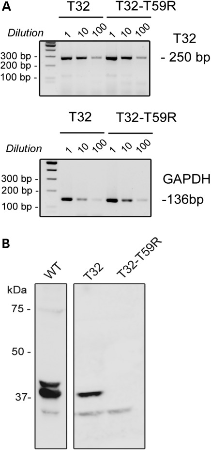
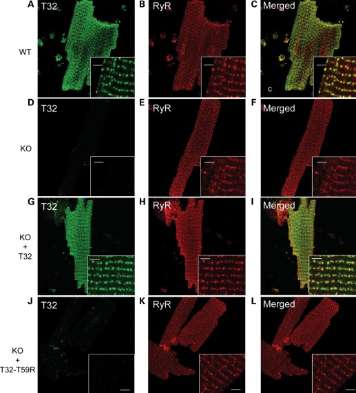
Catecholaminergic polymorphic ventricular tachycardia (CPVT) is an inherited arrhythmogenic disease so far related to mutations in the cardiac ryanodine receptor (RYR2) or the cardiac calsequestrin (CASQ2) genes. Because mutations in RYR2 or in CASQ2 are not retrieved in all CPVT cases, we searched for mutations in the physiological protein partners of RyR2 and CSQ2 in a large cohort of CPVT patients with no detected mutation in these two genes. Based on a candidate gene approach, we focused our investigations on triadin and junctin, two proteins that link RyR2 and CSQ2. Mutations in the triadin (TRDN) and in the junctin (ASPH) genes were searched in a cohort of 97 CPVT patients. We identified three mutations in triadin which cosegregated with the disease on a recessive mode of transmission in two families, but no mutation was found in junctin. Two TRDN mutations, a 4 bp deletion and a nonsense mutation, resulted in premature stop codons; the third mutation, a p.T59R missense mutation, was further studied. Expression of the p.T59R mutant in COS-7 cells resulted in intracellular retention and degradation of the mutant protein. This was confirmed after in vivo expression of the mutant triadin in triadin knock-out mice by viral transduction. In this work, we identified TRDN as a new gene responsible for an autosomal recessive form of CPVT. The mutations identified in the two families lead to the absence of the protein, thereby demonstrating the importance of triadin for the normal function of the cardiac calcium release complex in humans.


Figures

Figure 1.
Figure 1.
Triadin mutations in two CPVT families. (A) Pedigrees of the two CPVT families. Filled squares indicated affected individuals and half filled symbols individuals heterozygous for a mutation. The proband in each Family is indicated by the black arrow. The genotype for each individual concerning the identified mutation is indicated as follow: ‘+’ in the presence of the mutation and ‘−’ in the absence of the mutation. (B) Membrane topology of cardiac isoform of triadin, Trisk 32, in the sarcoplasmic reticulum membrane with the localization of the three identified mutations. The ‘KEKE’ region is the interaction domain with RyR2 and CSQ2. Trisk 32 has a C-terminal specific region from amino acid 264–286.
Figure 2.
Figure 2.
Trisk 32 and Trisk 32-T59R cell localizations are different. (A) Trisk 32-T59R is located only at the endoplasmic reticulum, whereas Trisk 32 is mainly at the plasma membrane. WT Trisk 32 (T32) and mutant Trisk 32-T59R (T32–T59R) were transfected in COS-7 cells, and their localization analyzed by immunofluorescent labeling. Typical Trisk 32 labeling at the plasma membrane (PM) or in the ER are shown. Bar: 20 µm. The histogram shows the percentage of transfected cells exhibiting PM (closed square) or ER (open square) labeling, for a total of 500 transfected cells from three different experiments. ***P < 0.001, Fisher's test comparison of T32- vs. T32–T59R-transfected cells. (B) Immunofluorescent labeling on intact or permeabilized cells shows intracellular retention of Trisk 32-T59R. Transfected cells were fixed without permeabilization and stained with an antibody directed against the C-terminal end of Trisk 32, which is extracellular when the protein is in the plasma membrane (left panels, ‘intact’). Afterwards, cells were permeabilized and stained with an antibody directed against the N-terminal end of Trisk 32, which is cytosolic when the protein is in the plasma membrane or in the reticulum membrane (right panels, ‘permeabilized’). Bar: 20 µm.
Figure 3.
Figure 3.
The mutant protein Trisk 32-T59R is degraded faster, via the proteasome. (A) Trisk 32-T59R is degraded faster in the course of protein synthesis inhibition. WT Trisk 32 (T32) and mutant Trisk 32-T59R (T32-T59R) were first transfected in COS-7 cells. Transfected cells were then incubated with cycloheximine (CHX) to block protein synthesis for the indicated time. Cells were collected and analyzed by quantitative western blot with specific antibodies to evaluate the amount of protein. The curves show the quantification from four experiments of the amount of each protein (T32 -•- and T32-T59R -□-) compared with its initial amount. ***P < 0.001, F-test comparison of the two curves, based on decay and plateau parameters. (B) Inhibition of the proteasome raises the amount of the mutant protein Trisk 32-T59R. Twenty hours after transfection, the cells were incubated during 6 h with either dimethyl sulfoxide (DMSO) alone for controls (lanes ‘−’) or 50 µm MG132 to block proteasome (lanes ‘+’). The cells were then collected and analyzed by quantitative western blot with specific antibodies. The top panel shows a typical western blot, the bottom histogram (▪ DMSO; □ MG132) is a quantification (mean ± SEM) from three experiments. *P < 0.05, Mann and Whitney test comparison of DMSO- vs. MG132-treated cells.
Figure 4.
Figure 4.
Stability analyses of transcripts and proteins after in vivo expression in triadin KO mice. (A) The mutant transcript is present at similar levels as the WT one. Messenger RNAs were purified from isolated cardiomyocytes of transduced mice. After reverse transcription, the cDNAs were either directly used or diluted 10 or 100 times before the PCR amplification of Trisk 32 (T32) and GAPDH. PCR products for Trisk 32 and GAPDH amplification were expected, respectively, at 250 and 136 bp. The PCR primers for GAPDH were designed to co-amplify possible contaminating genomic DNA as a 220 bp fragment, which was not observed. (B) The mutant protein Trisk 32-T59R was not detectable. Western blot analysis of 100 µg of cardiomyocytes homogenates from control WT mouse, with mouse-specific anti-Trisk 32 antibody (first lane), or 100 µg of cardiomyocytes from a Trisk 32-transduced mouse (second lane), or Trisk 32-T59R-transduced mouse (third lane) with rat-specific anti-Trisk 32 antibody. Trisk 32 appears as multiple bands (2/3) centered on 37 kDa, the higher bands, the glycosylated forms of Trisk 32, being in lower amount and visible in correlation with the intensity of the signal, as described before (6,20).
Figure 5.
Figure 5.
Immunofluorescent analysis of Trisk 32 in isolated cardiomyocytes. Cardiomyocytes were isolated from a WT mouse, a triadin KO mouse (KO), a triadin KO mouse transduced with rat Trisk 32 (KO + T32) and a triadin KO mouse transduced with rat Trisk 32-T59R (KO + T32-T59R). They were labeled with antibodies against mouse Trisk 32 and RyR (AF) or against rat Trisk 32 and RyR (GL). In all cardiomyocytes, RyR labeling is typical of a dyad labeling showing aligned rows of dots, as observed in the inserts. Bar: 2 µm.

References

    1. Hayashi M., Denjoy I., Extramiana F., Maltret A., Buisson N.R., Lupoglazoff J.M., Klug D., Hayashi M., Takatsuki S., Villain E., et al. Incidence and risk factors of arrhythmic events in catecholaminergic polymorphic ventricular tachycardia. Circulation. 2009;119:2426–2434. doi:10.1161/CIRCULATIONAHA.108.829267. - DOI - PubMed
    1. Priori S.G., Chen S.R. Inherited dysfunction of sarcoplasmic reticulum Ca2+ handling and arrhythmogenesis. Circ. Res. 2011;108:871–883. doi:10.1161/CIRCRESAHA.110.226845. - DOI - PMC - PubMed
    1. Guo W., Campbell K.P. Association of triadin with the ryanodine receptor and calsequestrin in the lumen of the sarcoplasmic reticulum. J. Biol. Chem. 1995;270:9027–9030. doi:10.1074/jbc.270.16.9027. - DOI - PubMed
    1. Zhang L., Kelley J., Schmeisser G., Kobayashi Y.M., Jones L.R. Complex formation between junctin, triadin, calsequestrin, and the ryanodine receptor. Proteins of the cardiac junctional sarcoplasmic reticulum membrane. J. Biol. Chem. 1997;272:23389–23397. doi:10.1074/jbc.272.37.23389. - DOI - PubMed
    1. Thevenon D., Smida-Rezgui S., Chevessier F., Groh S., Henry-Berger J., Romero N.B., Villaz M., De Waard M., Marty I. Human skeletal muscle triadin: gene organization and cloning of the major isoform, Trisk 51. Biochem. Biophys. Res. Commun. 2003;303:669–675. doi:10.1016/S0006-291X(03)00406-6. - DOI - PubMed

Publication types

MeSH terms