. 2012 Apr 15;188(8):4113-21.
doi: 10.4049/jimmunol.1103113. Epub 2012 Mar 14.

Pleiotropic IFN-dependent and -independent effects of IRF5 on the pathogenesis of experimental lupus

Affiliations

Pleiotropic IFN-dependent and -independent effects of IRF5 on the pathogenesis of experimental lupus

Yuan Xu et al. J Immunol. .

Abstract

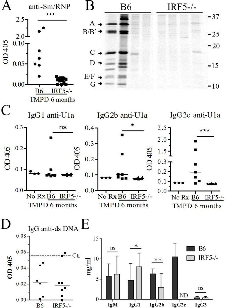
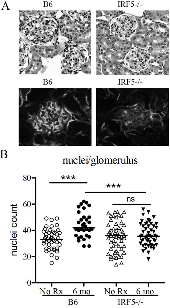
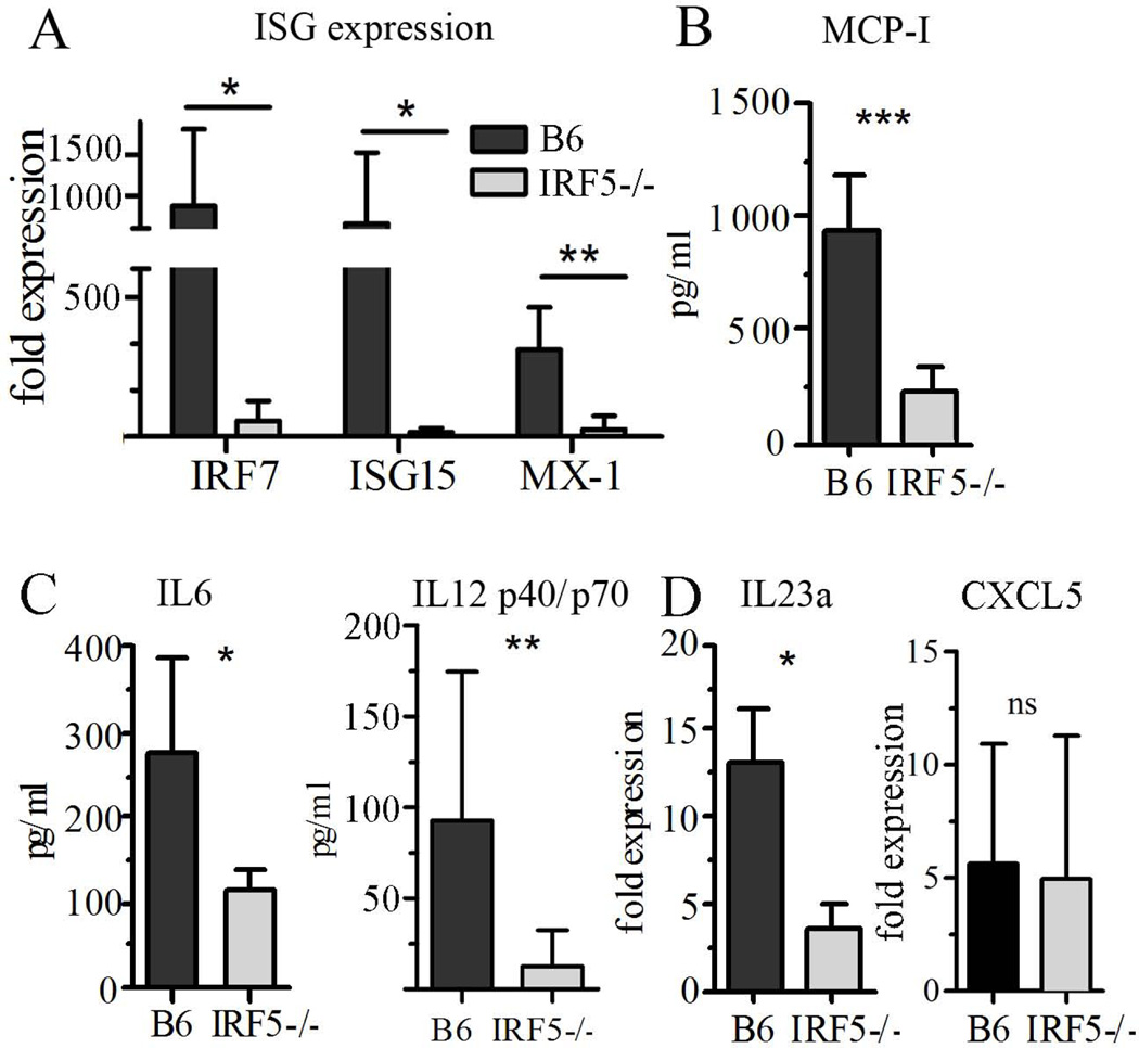
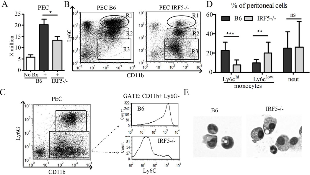
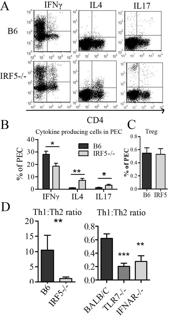
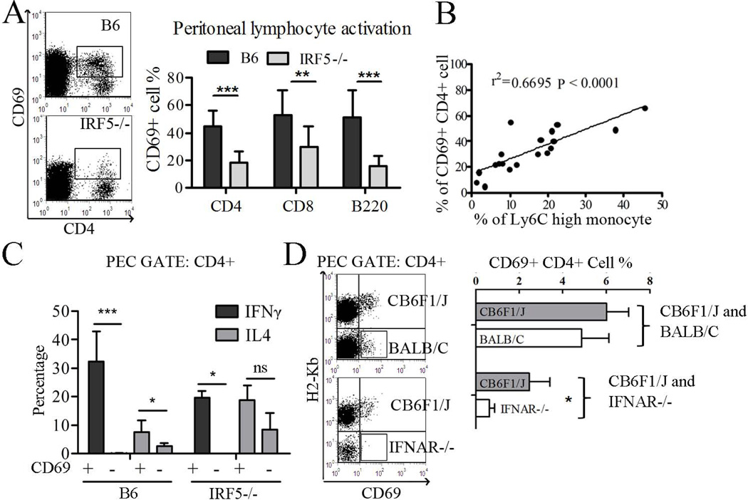
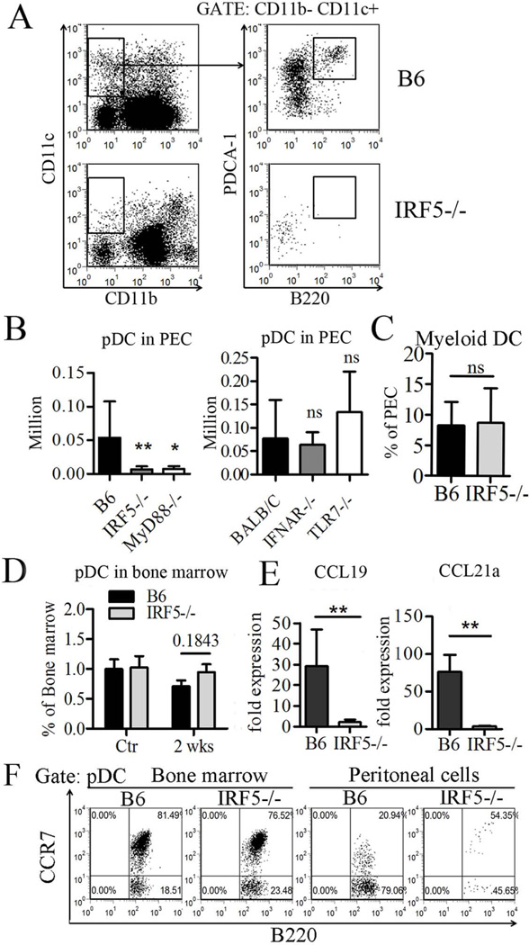
Genetic polymorphisms of IFN regulatory factor 5 (IRF5) are associated with an increased risk of lupus in humans. In this study, we examined the role of IRF5 in the pathogenesis of pristane-induced lupus in mice. The pathological response to pristane in IRF5(-/-) mice shared many features with type I IFN receptor (IFNAR)(-/-) and TLR7(-/-) mice: production of anti-Sm/RNP autoantibodies, glomerulonephritis, generation of Ly6C(hi) monocytes, and IFN-I production all were greatly attenuated. Lymphocyte activation following pristane injection was greatly diminished in IRF5(-/-) mice, and Th cell differentiation was deviated from Th1 in wild-type mice toward Th2 in IRF5(-/-) mice. Th cell development was skewed similarly in TLR7(-/-) or IFNAR(-/-) mice, suggesting that IRF5 alters T cell activation and differentiation by affecting cytokine production. Indeed, production of IFN-I, IL-12, and IL-23 in response to pristane was markedly decreased, whereas IL-4 increased. Unexpectedly, plasmacytoid dendritic cells (pDC) were not recruited to the site of inflammation in IRF5(-/-) or MyD88(-/-) mice, but were recruited normally in IFNAR(-/-) and TLR7(-/-) mice. In striking contrast to wild-type mice, pristane did not stimulate local expression of CCL19 and CCL21 in IRF5(-/-) mice, suggesting that IRF5 regulates chemokine-mediated pDC migration independently of its effects on IFN-I. Collectively, these data indicate that altered production of IFN-I and other cytokines in IRF5(-/-) mice prevents pristane from inducing lupus pathology by broadly affecting T and B lymphocyte activation/differentiation. Additionally, we uncovered a new, IFN-I-independent role of IRF5 in regulating chemokines involved in the homing of pDCs and certain lymphocyte subsets.


Figures

Figure 1
Figure 1. Absence of autoantibodies in IRF5−/− mice
IRF5−/− and control B6 mice (B6 mice N=7, IRF5−/− mice N=14) were treated with pristane and 6 months later, serum autoantibody and immunoglobulin levels were evaluated. Sera collected from mice before pristane (tetramethylpentadecane, TMPD) injection served as a control (Ctr). A, Anti-Sm/RNP autoantibody levels (ELISA) in IRF5−/− and B6 control mice. B, immunoprecipitation of [35S]-radiolabeled protein components A–G of U1 small ribonucleoproteins by sera from 6 B6 and 6 IRF5−/− sera. Positions of molecular weight markers (kDa) are indicated on the right. C, Anti-U1A IgG1, IgG2b and IgG2c autoantibody levels (ELISA) in IRF5−/− and control mice (B6 mice N=7, IRF5−/− mice N=14). D, IgG anti-dsDNA autoantibody levels (ELISA) in IRF5−/− and control mice (B6 mice N=6, IRF5−/− mice N=8). Dotted line indicates the mean absorbance using sera from non-pristane-treated control mice. E, Serum levels of total IgM, IgG1, IgG2c, IgG2b, and IgG3 in pristane-treated IRF5−/− and B6 mice (B6 mice N=7, IRF5−/− mice N=14). ND: not detected. * P < 0.05; ** P <0.01; *** P <0.001 by Mann-Whitney test (Figure A, C, D, E).
Figure 2
Figure 2. Attenuated renal disease in pristane-treated IRF5−/− mice
A, Upper: kidneys from IRF5−/− mice and wild type B6 controls 6 months after pristane treatment were sectioned and stained with hematoxylin & eosin. Lower: Renal tissue was embedded in OCT and 4 µm sections were stained with FITC-conjugated anti-mouse complement component C3 monoclonal antibody. B, Glomerular cellularity was evaluated by counting the number of nuclei per glomerular cross-section (n=5 mice per group, 8 glomeruli per mouse). *** P <0.001, Student’s t-test.
Figure 3
Figure 3. Decreased levels of inflammatory cytokines and chemokines in IRF5−/− mice
IRF5−/− and B6 mice were injected with pristane and 2–3 weeks later, the peritoneum was lavaged and peritoneal exudate cells were collected by centrifugation for RNA isolation. The supernatant was used to assay cytokine/chemokine levels. A, Expression of the interferon-stimulated genes (ISGs) IRF7, ISG15, and Mx-1 in peritoneal exudate cells (Q-PCR). B, Level of MCP-1 (CCL2) in peritoneal lavage fluid (ELISA). C, Levels of IL-6 and IL-12 P40/P70 in peritoneal lavage fluid (ELISA). D, Expression of IL-23a (p19) and CXCL5 in peritoneal exudate cells (Q-PCR). * P <0.05; ** P <0.01; *** P <0.001 by Mann-Whitney test.
Figure 4
Figure 4. Effect of IRF5 deficiency on the inflammatory response to pristane
Two weeks after pristane injection, peritoneal cells were collected from IRF5−/− and wild type (B6) controls and analyzed by flow cytometry. A, Total peritoneal exudate cell count. B, Analysis of total peritoneal exudate cells by flow cytometry. Percentages of CD11b+Ly6Chi monocytes (R1), CD11b+Ly6Cint neutrophils (R2), and CD11b+Ly6Clo “mature” monocytes (R3) were assessed by flow cytometry as described previously (29). C, Fluorescence intensity of Ly6C on CD11b+Ly6G monocytes in the peritoneal exudates was assessed by flow cytometry. D, percentages of Ly6Chi monocytes (CD11b+Ly6ChiLy6G), Ly6Clo monocytes (CD11b+Ly6CloLy6G), and neutrophils (CD11b+Ly6CintLy6G+) in the peritoneal exudates. Quantification was based on panel C. E, Morphology of peritoneal exudate monocytes from pristane-treated wild type (B6) vs. IRF5−/− mice. N = 14–22 per group, representative of two independent experiments. * P < 0.05; ** P <0.01; *** P <0.001 by Mann-Whitney test.
Figure 5
Figure 5. Inflammatory response to pristane is skewed toward TH2 cells IRF5−/− mice
Two weeks after pristane injection, peritoneal lymphocytes were analyzed by flow cytometry. A, flow cytometry of IFNγ+, IL4+, and IL17+ cells in total peritoneal exudate cells from IRF5−/− vs. B6 control mice. B, Quantification of IFNγ, IL4, and IL17 producing peritoneal cells. C, Quantification of CD4+FoxP3+ (Treg-like) cells in IRF5−/− vs. B6 mice. D, TH1/TH2 ratio in peritoneal exudates of IRF5−/− vs. B6 mice and TLR7−/−, IFNAR−/−, and wild type BALB/c mice 2 weeks after pristane injection. Representative of two independent experiments, n = 6–14 mice per group, * P < 0.05; ** P <0.01; *** P <0.001 by Mann-Whitney test.
Figure 6
Figure 6. Role of IRF-5 in lymphocyte activation following pristane treatment
Two weeks after pristane injection, peritoneal lymphocytes were analyzed by flow cytometry. A, Left: flow cytometry of CD4 and CD69 (gated on CD11b cells). Right: The percentage of CD4+, CD8+, and B220+ cells that were positive for CD69 was determined. B, Correlation of CD69+ CD4+ T cells with the percentage of Ly6Chi monocytes. C, Intracellular staining of IFNγ and IL4 in B6 and IRF5−/− mice. Peritoneal lymphocytes were gated on the CD4+ population and then gated on the CD69+ and CD69 populations, respectively. Percentages of cells expressing IFNγ and IL4 are indicated (N=4 per group). ND, not detected. D, Left: peritoneal cells from bone marrow chimeric mice were gated on the CD11bCD4+ population and CD69 expression was analyzed by flow cytometry in cells that were H2-Kb+ (CB6F1/J derived) or H2-Kb− (BALB/c or BALB/c IFNAR−/− derived). Right, quantification of CD69+CD4+ T cells as a percentage of total CD4+ T cells (N=4–5 per group). PEC, peritoneal exudate cells. Data are representative of two independent experiments. * P < 0.05; ** P <0.01; *** P <0.001 by Mann-Whitney test.
Figure 7
Figure 7. Decreased plasmacytoid dendritic cells (pDCs) in the peritoneum of IRF5−/−, but not TLR7−/− or IFNAR−/−, mice
Two weeks after pristane injection, peritoneal exudate and bone marrow pDCs were quantified by flow cytometry. A, pDC staining. CD11b CD11c+B220+PDCA-1+ cells were defined as pDC; this cell population was also Ly6C+ (not shown). B, Left, pDCs in peritoneal exudate cells (PEC) from IRF5−/−, MyD88−/−, and B6 control mice (N=5–12 per group). Right, pDC in TLR7−/−, IFNAR−/− and BALB/cJ PEC (N=6 per group). C, Peritoneal myeloid dendritic cells (CD11b+CD11c+CD8) were quantified by flow cytometry (N=9–10 mice per group). D, pDCs in bone marrow of control B6 IRF5−/− and B6 mice before pristane injection (Ctr) and 2 weeks after pristane injection (N=3–5 per group). E, Expression of chemokines CCL19 and CCL21a in PEC by Q-PCR (N=4–6 per group). F, Expression of CCR7 on bone marrow and peritoneal pDCs (CD11b CD11c+B220+PDCA-1+) was determined by flow cytometry (N=4 per group). Data are representative of two independent experiments. * P < 0.05; ** P <0.01; *** P <0.001 Mann-Whitney test.

References

    1. Takaoka A, Yanai H, Kondo S, Duncan G, Negishi H, Mizutani T, Kano S, Honda K, Ohba Y, Mak TW, Taniguchi T. Integral role of IRF-5 in the gene induction programme activated by Toll-like receptors. Nature. 2005;434:243–249. - PubMed
    1. Barnes BJ, Moore PA, Pitha PM. Virus-specific activation of a novel interferon regulatory factor, IRF-5, results in the induction of distinct interferon alpha genes. J Biol Chem. 2001;276:23382–23390. - PubMed
    1. Schoenemeyer A, Barnes BJ, Mancl ME, Latz E, Goutagny N, Pitha PM, Fitzgerald KA, Golenbock DT. The interferon regulatory factor, IRF5, is a central mediator of toll-like receptor 7 signaling. J Biol Chem. 2005;280:17005–17012. - PubMed
    1. Couzinet A, Tamura K, Chen HM, Nishimura K, Wang Z, Morishita Y, Takeda K, Yagita H, Yanai H, Taniguchi T, Tamura T. A cell-type-specific requirement for IFN regulatory factor 5 (IRF5) in Fas-induced apoptosis. Proc Natl Acad Sci U S A. 2008;105:2556–2561. - PMC - PubMed
    1. Lien C, Fang CM, Huso D, Livak F, Lu R, Pitha PM. Critical role of IRF-5 in regulation of B-cell differentiation. Proc Natl Acad Sci U S A. 2010;107:4664–4668. - PMC - PubMed

Publication types

MeSH terms