. 2012 Mar 16;335(6074):1366-9.
doi: 10.1126/science.1217039.

Decoding in the absence of a codon by tmRNA and SmpB in the ribosome

Affiliations

Decoding in the absence of a codon by tmRNA and SmpB in the ribosome

Cajetan Neubauer et al. Science. .

Abstract

In bacteria, ribosomes stalled at the end of truncated messages are rescued by transfer-messenger RNA (tmRNA), a bifunctional molecule that acts as both a transfer RNA (tRNA) and a messenger RNA (mRNA), and SmpB, a small protein that works in concert with tmRNA. Here, we present the crystal structure of a tmRNA fragment, SmpB and elongation factor Tu bound to the ribosome at 3.2 angstroms resolution. The structure shows how SmpB plays the role of both the anticodon loop of tRNA and portions of mRNA to facilitate decoding in the absence of an mRNA codon in the A site of the ribosome and explains why the tmRNA-SmpB system does not interfere with normal translation.


Figures
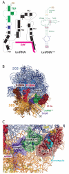
Fig. 1
Fig. 1
Overview of the structure of Ala-tmRNAΔm, SmpB and EF-Tu·GDP bound to the ribosome. (A) Secondary structure diagram of tmRNA and sequence of the tmRNAΔm fragment used in this study. The tRNA-like domain (TLD) is highlighted in green, the open reading frame (ORF) in magenta and RNA helix 2b in blue. Mutations introduced for improved refolding of the in vitro transcribed RNA and the UUCG tetra loop are indicated in magenta, the G·U base pair recognized by alanine tRNA synthetase in yellow. (B) Overview of the ribosomal complex with EF-Tu, SmpB and tmRNAΔm. (C) Detailed view of the ribosomal A site.
Fig. 2
Fig. 2
Superposition of tmRNAΔm·SmpB·EF-Tu with a complex of A-site tRNA and EF-Tu (gray, PDB code 2Y10, ref. 36). In the tmRNA·SmpB complex, SmpB substitutes for the anticodon arm of tRNA. The interactions of EF-Tu (red) and the acceptor-stem region of tmRNA (green) are nearly identical to that observed for the tRNA-EF-Tu complex (gray). A close-up view of the 3′-end CCA sequence is shown as inset.
Fig. 3
Fig. 3
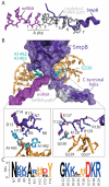
Interactions of SmpB with the ribosome. (A) The C-terminal tail of SmpB would clash with mRNA downstream of the A-site codon. The mRNA used in this work is colored in magenta and an extension based on the superposition of a longer mRNA (PDB code 2HGR, ref. 31) is shown in gray. The mRNA nucleotides are numbered starting with the first nucleotide of the A-site codon. (B) SmpB interacts with both the shoulder domain and the 3′ major domain of 16S rRNA near the decoding center. Close-up views of the interactions near A1492/A1493 and G530 at the decoding center are shown in insets. (C) Sequence alignment for the regions of SmpB interacting with the decoding center with degree of conservation indicated by size. Positively charged residues are highlighted in blue, negative in red and aromatic in yellow. See SOM and Fig. S4 for details.

References

    1. Keiler KC, Waller PR, Sauer RT. Science. 1996;271:990. - PubMed
    1. Moore SD, Sauer RT. Annu Rev Biochem. 2007;76:101. - PubMed
    1. Komine Y, Kitabatake M, Yokogawa T, Nishikawa K, Inokuchi H. Proc Natl Acad Sci U S A. 1994;91:9223. - PMC - PubMed
    1. Martin A, Baker TA, Sauer RT. Mol Cell. 2008;29:441. - PMC - PubMed
    1. Karzai AW, Susskind MM, Sauer RT. EMBO J. 1999;18:3793. - PMC - PubMed

Publication types

MeSH terms