. 2012 Mar 16;335(6074):1370-2.
doi: 10.1126/science.1217443.

Structural basis for the rescue of stalled ribosomes: structure of YaeJ bound to the ribosome

Affiliations

Structural basis for the rescue of stalled ribosomes: structure of YaeJ bound to the ribosome

Matthieu G Gagnon et al. Science. .

Abstract

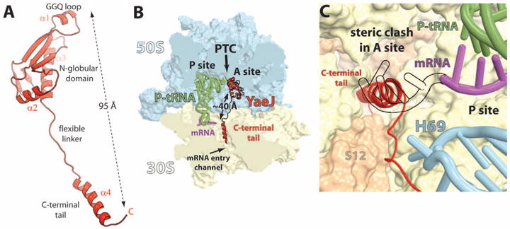
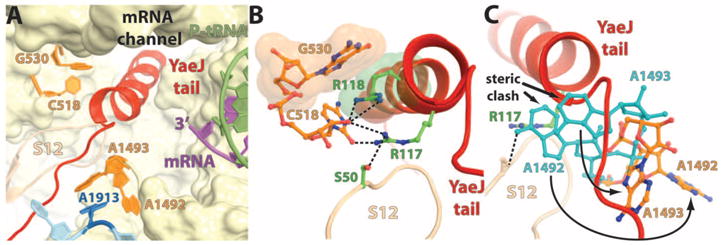
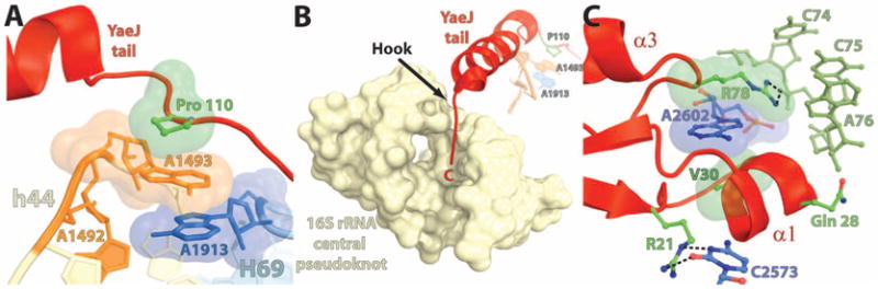
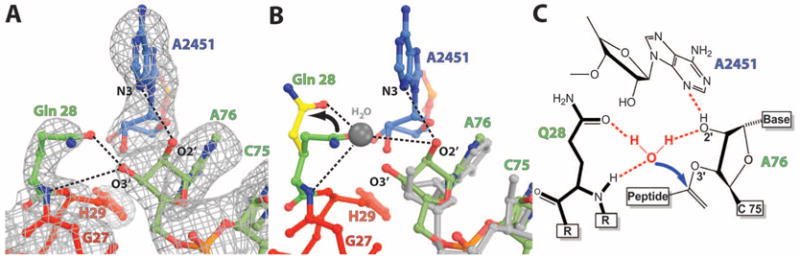
In bacteria, the hybrid transfer-messenger RNA (tmRNA) rescues ribosomes stalled on defective messenger RNAs (mRNAs). However, certain gram-negative bacteria have evolved proteins that are capable of rescuing stalled ribosomes in a tmRNA-independent manner. Here, we report a 3.2 angstrom-resolution crystal structure of the rescue factor YaeJ bound to the Thermus thermophilus 70S ribosome in complex with the initiator tRNA(i)(fMet) and a short mRNA. The structure reveals that the C-terminal tail of YaeJ functions as a sensor to discriminate between stalled and actively translating ribosomes by binding in the mRNA entry channel downstream of the A site between the head and shoulder of the 30S subunit. This allows the N-terminal globular domain to sample different conformations, so that its conserved GGQ motif is optimally positioned to catalyze the hydrolysis of peptidyl-tRNA. This structure gives insights into the mechanism of YaeJ function and provides a basis for understanding how it rescues stalled ribosomes.


Figures

Fig. 1
Fig. 1
The structure of YaeJ bound to the ribosome. (A) Cartoon representation of YaeJ shown in its ribosome-bound conformation. (B) Overview of YaeJ, P-site tRNA, and mRNA bound to the 70S ribosome. The 50S and 30S subunits are shown in light blue and yellow, respectively. Portions of the ribosome are omitted for clarity. The N-terminal domain of YaeJ is bound in the A site of the 50S subunit, adjacent to the CCA end of the peptidyl-tRNA, and the C-terminal tail occupies the mRNA entry channel in the 30S. (C) Positioning of the α-helical YaeJ tail in the mRNA entry channel. The mRNA in the present complex (magenta) ends in the P site. The mRNA making codon-anticodon interaction in the A site (shown as a black outline) [2J00, (15)] was superimposed on our model to reveal the steric clash with the YaeJ tail.
Fig. 2
Fig. 2
Interactions of the YaeJ tail with the universally conserved nucleotides in the decoding center. (A) Overview of the YaeJ tail in the decoding center and its interaction with the key bases that are labeled. (B) Close-up view of the stacking between G530 (16S rRNA) and R118 of YaeJ shown as space-filling representation in orange and green, respectively. Putative hydrogen bonds of R117 with C518 (16S rRNA) and S50 from ribosomal protein S12 are shown as black dashes. (C) Conformational differences between A1492 and A1493 from our model (shown in orange) relative to their counterparts from a structure with an A site–bound tRNA [2J00 (15)] (shown in light blue) reveal that the latter sterically clash with the YaeJ tail. The positioning of these key bases in the present complex is indicated by the curved arrows.
Fig. 3
Fig. 3
Stabilization of the YaeJ tail in the mRNA entry channel (A and B) and the GGQ loop in the PTC (C). (A) Stacking between Pro110 (P110) from YaeJ (green), A1493 from h44 (orange), and A1913 from H69 (blue). (B) Interaction of the C-terminal end of YaeJ (residues 129 to 133) with the 16S rRNA central pseudoknot consisting of helix 1 (nucleotides 9 to 13 and 21 to 25) and helix 2 (nucleotides 17 to 19 and 916 to 918). A1493 from h44 in the decoding center (orange), A1913 from H69 (blue), and P110 from YaeJ (green) are shown in the background. The interactions shown in (A) and (B) together anchor the YaeJ tail in the mRNA entry channel. (C) The potential hydrogen bonds are shown as black dashes. The H-bonds between C2573 and R21, R78, and C75 of the P-tRNA and the stacking between A2602, R78, and V30 (shown as space-filling representation) help to position the GGQ loop next to the CCA end of the P-tRNA.
Fig. 4
Fig. 4
Positioning of the GGQ loop and catalysis. (A) σA-weighted 2Fob sFcalc electron density map contoured at 1.2σ showing the interactions of the GGQ loop at the site of catalysis. Putative hydrogen bonds between Gln28, A2451, and the riboseofA76 are shownasblack dashes. (B) Proposed catalytic mechanism reveals that a small change in dihedral angles of Gln28 as indicated by the arrow (green versus yellow) allows it to coordinate the nucleophilic water [shown as a gray ball modeled based on 1VQ7 (25)] that attacks the aminoacyl ester bond. (C) Schematic representation of the proposed catalysis reaction shown in (B).

References

    1. Felden B, Gillet R. RNA Biol. 2011;8:440. - PubMed
    1. Keiler KC, Waller PR, Sauer RT. Science. 1996;271:990. - PubMed
    1. Withey J, Friedman D. J Bacteriol. 1999;181:2148. - PMC - PubMed
    1. Handa Y, Inaho N, Nameki N. Nucleic Acids Res. 2011;39:1739. - PMC - PubMed
    1. Garza-Sánchez F, Schaub RE, Janssen BD, Hayes CS. Mol Microbiol. 2011;80:1204. - PMC - PubMed

Publication types

MeSH terms