. 2012 Mar 14;4(125):125ra30.
doi: 10.1126/scitranslmed.3003481.

A model for personalized in vivo analysis of human immune responsiveness

Affiliations

A model for personalized in vivo analysis of human immune responsiveness

Hannes Kalscheuer et al. Sci Transl Med. .

Abstract

Studies of human immune diseases are generally limited to the analysis of peripheral blood lymphocytes of heterogeneous patient populations. Improved models are needed to allow analysis of fundamental immunologic abnormalities predisposing to disease and in which to assess immunotherapies. Immunodeficient mice receiving human fetal thymus grafts and fetal CD34(+) cells intravenously produce robust human immune systems, allowing analysis of human T cell development and function. However, to use humanized mice to study human immune-mediated disorders, immune systems must be generated from adult hematopoietic cells. Here, we demonstrated robust immune reconstitution in mice with hematopoietic stem cells (HSCs) aspirated from bone marrow of adults with type 1 diabetes (T1D) and healthy control volunteers. In these humanized mice, cryopreservation of human leukocyte antigen allele-matched fetal thymic tissue prevented allogeneic adult HSC rejection. Newly generated T cells, which included regulatory T cells (T(regs)), were functional and self-tolerant and had a diverse repertoire. The immune recognition of these mice mimicked that of the adult CD34(+) cell donor, but the T cell phenotypes were more predominantly "naïve" than those of the adult donors. HSCs from T1D and control donors generated similar numbers of natural T(regs) intrathymically; however, peripheral T cells from T1D subjects showed increased proportions of activated or memory cells compared to controls, suggesting possible HSC-intrinsic differences in T cell homeostasis that might underlie immune pathology in T1D. This "personalized immune" mouse provides a new model for individualized analysis of human immune responses that may provide new insights into not only T1D but also other forms of immune function and dysfunction as well.


Conflict of interest statement

The authors declare that they have no competing interests.

Figures
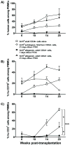
Figure 1
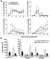
Figure 1. Peripheral human cell reconstitution in NOD/SCID mice following transplantation of dGuo-treated thymic tissue
Fetal thymic tissue was treated with dGuo for 7 (black diamonds, n=4) or 21 days (black triangles, n=5) before transplantation into sublethally irradiated NOD/SCID that received 5×10⁵ allogeneic, adult CD34+ cells or 4×10⁵ autologous fetal liver CD34+ cells (open circles, n=5) intravenously. Age-matched control animals received 5×10⁵ adult CD34+ cells alone (open squares, n=5). The mice were bled to measure human (hu) cell reconstitution in (total mouse plus human) peripheral blood mononuclear cells (PBMCs) at the indicated time points. A: total human chimerism; B: percentage of human B cells; C: percentage of human T cells among PBMCs at indicated time points.
Figure 2
Figure 2. Multilineage human cell reconstitution in NSG mice receiving cryopreserved and thawed thymic grafts and allogeneic, adult CD34+ cells
A) Sublethally irradiated NSG mice that received a cryopreserved and thawed fetal thymus graft in combination with 3×10⁵ (black squares, n= 6) or 5×10⁵ (black triangles, n=6) adult CD34+ cells and two doses of anti-CD2 mAb i.v. were bled to measure human cell reconstitution in (total mouse plus human) PBMCs at the indicated time points. Age-matched control animals received 3×10⁵ adult HSCs alone (white squares, n=6). Single cell suspensions of PBMCs were stained for markers of human hematopoietic cells (CD45), T cells (CD3), B cells (CD19) and monocytes (CD14). Dead cells and mouse red blood cells were excluded from the analysis. B) Representative THY graft appearance 20 weeks post-transplantation. NSG mice that were transplanted with cryopreserved and thawed fetal thymus tissue had abundant, viable thymic tissue underneath the kidney capsule. C) FCM analysis of thymocytes (representative of 12 grafts in a single experiment).
Figure 3
Figure 3. Multilineage human cell reconstitution in NSG mice receiving cryopreserved/thawed thymic grafts and allogeneic, adult CD34+ cells isolated from bedside bone marrow aspirates
A) Sublethally irradiated NSG mice received cryopreserved/thawed fetal thymus tissue in combination with 1.8-3.0×10⁵ adult CD34+ cells isolated from bone marrow aspirates from healthy volunteers (black squares, n=3 donors, 6 recipients) and T1D subjects (black triangles, n=4 donors, 29 recipients). Mean levels of human cell reconstitution in (total mouse plus human) PBMCs are shown over time. Control animals received adult HSCs alone from the T1D subjects (open circles, n=2 donors, 7 recipients). Thymus grafts and bone marrow donors were HLA-typed for T1D-associated DRB and DQB alleles and HLA A*201 using SNP genotyping assays. The thymic tissue and bone marrow donors shared at least HLA*A201 and DRB*0302 and/or DQB*0301. ANOVA revealed an effect of THY transplant on CD3 reconstitution comparing T1D CD34+ cells alone with T1D CD34+ plus THY transplantation at early (6-14 weeks) timepoints. Individual timepoint comparisions with Mann-Whitney U test revealed significant differences at 8-14 weeks (*p<0.05). B) Graft thymocytes were analyzed 22-25 weeks post-transplantation in T1D (n=5) and control (n=3) HSC-reconstituted animals. Mean + SEM are shown. No significant differences between T1D and control animals were noted.
Figure 4
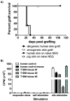
Figure 4. Functional and self-tolerant immune systems in NSG mice receiving fetal thymus graft and adult CD34+ cells
A) NSG mice (n=3) that received a 7 Gy irradiated thymic graft plus 3×10⁵ adult CD34+ cells reconstituted peripheral T cells >30 weeks after transplantation. Thirty-nine weeks after transplantation, they were grafted with allogeneic human and xenogeneic pig skin. Survival of pig and human skin grafts (n=3 and 4, respectively) on untreated control NSG mice “naïve NSG” are also shown. B) Human T cells (>90% pure) were enriched from the spleen and peripheral lymph nodes of NSG mice 20 weeks following transplantation of cryopreserved and thawed THY grafts and allogeneic bone marrow CD34+ cells from a healthy control (black bar) or T1D subject (dotted bars). Control T cells were isolated from PBMC of the same healthy control volunteer (open bar). Supplementary Table 1 shows the naïve/memory cell distribution of CD4 cells in the 3 mice reconstituted from T1D CD34+ cells.
Figure 5
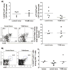
Figure 5. Tregs in thymus grafts and periphery of PI mice
A) 20-22 weeks after transplantation, single cell suspensions were prepared from thymus grafts of NSG mice that received a cryopreserved and thawed THY graft and allogeneic CD34+ cells from one of two healthy volunteers (circles) or one T1D subject (squares) and analysed for CD4+CD8-CD25+FoxP3+ cells by FCM (top row). As a marker for natural Tregs, Helios expression in CD4+CD8-CD25+CD127loFoxP3+ thymocytes is shown in NSG mice derived from a second human donor pair in the bottom row. B) Similar proportions of Tregs in PBMCs 20 weeks after transplantation of cryopreserved/thawed fetal thymus grafts with CD34+ cells from one of two healthy controls (black squares) or one T1D subject (black triangles) compared to two healthy humans (black circles). Left plots show CD25 and FoxP3 staining on CD4+ T cells from PI mice generated from control and T1D donors.
Figure 6
Figure 6. Diverse repertoire of T cells in PI mice
Spectratyping (β-chain CDR3 length distribution) of human CD4 and CD8 SP T cells in THY grafts reconstituted with CD34+ cells from one T1D donor or one of two healthy controls 20 weeks after transplantation. Spectratype from one representative animal (#5700) of six (mice) is shown. The vertical axis is relative fluorescence units (full scale=6,000 units). The horizontal axis is nucleotide size. Reference size markers are low fainter peaks. Representative BV is shown from a total of 12 analyzed per sample. The Hamming distances of all 6 samples, a measure of the relative distances of the observed TCR β-chain length distribution from a reference distribution of healthy adult CD4 T cells, are shown in Supplemental Table 1, and each indicates the reconstitution of a polyclonal repertoire.
Figure 7
Figure 7. Naïve vs memory phenotype of T cells in PI mice
A) and B) Proportions of CD45RA+ CD4 and CD8 T cells (B) and Tregs (C) in PBMCs of healthy volunteers and of PI mice 20 weeks after THY implantation plus i.v. infusion of CD34+ cells from one T1D subject (black squares) or one of two healthy controls (black triangles), including (open circle) the donor of CD34+ cells for the control mouse indicated with an open triangle (*p<0.05, excluding the outlier in the CD8 population of controls from statistical analysis). C) T cell populations were assayed by FCM in NSG mice injected with CD34+ fetal liver cells with or without allogeneic thymus at 7 weeks post-transplant. Mean ± SEM are shown, n=4 for each group. *p<0.05, **p<0.005.

References

    1. Mosier DE, Gulizia RJ, Baird SM, Wilson DB. Transfer of a functional human immune system to mice with severe combined immunodeficiency. Nature. 1988;335:256–259. - PubMed
    1. Tary-Lehmann M, Lehmann P, Schols D, Roncarolo MG, Saxon A. Anti-SCID mouse reactivity shapes the human CD4+ T cell repertoire in hu-PBL-SCID chimeras. J Exp Med. 1994;180:1817–1827. - PMC - PubMed
    1. McCune JM, Namikawa R, Kaneshima H, Shultz LD, Lieberman M, Weissman IL. The SCID-hu mouse: Murine model for the analysis of human hematolymphoid differentiation and function. Science. 1988;241:1632–1639. - PubMed
    1. Lan P, Tonomura N, Shimizu A, Wang S, Yang YG. Reconstitution of a functional human immune system in immunodeficient mice through combined human fetal thymus/liver and CD34+ cell transplantation. Blood. 2006;108:487–492. - PubMed
    1. Tonomura N, Habiro K, Shimizu A, Sykes M, Yang YG. Antigen-specific human T-cell responses and T cell-dependent production of human antibodies in a humanized mouse model. Blood. 2008;111:4293–4296. - PMC - PubMed

Publication types