Comparative Study
. 2012 Mar 14;32(11):3818-29.
doi: 10.1523/JNEUROSCI.5775-11.2012.

Motor neuron rescue in spinal muscular atrophy mice demonstrates that sensory-motor defects are a consequence, not a cause, of motor neuron dysfunction

Affiliations
Comparative Study

Motor neuron rescue in spinal muscular atrophy mice demonstrates that sensory-motor defects are a consequence, not a cause, of motor neuron dysfunction

Rocky G Gogliotti et al. J Neurosci. .

Abstract

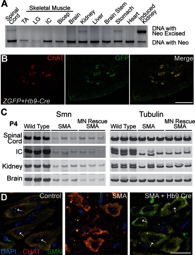
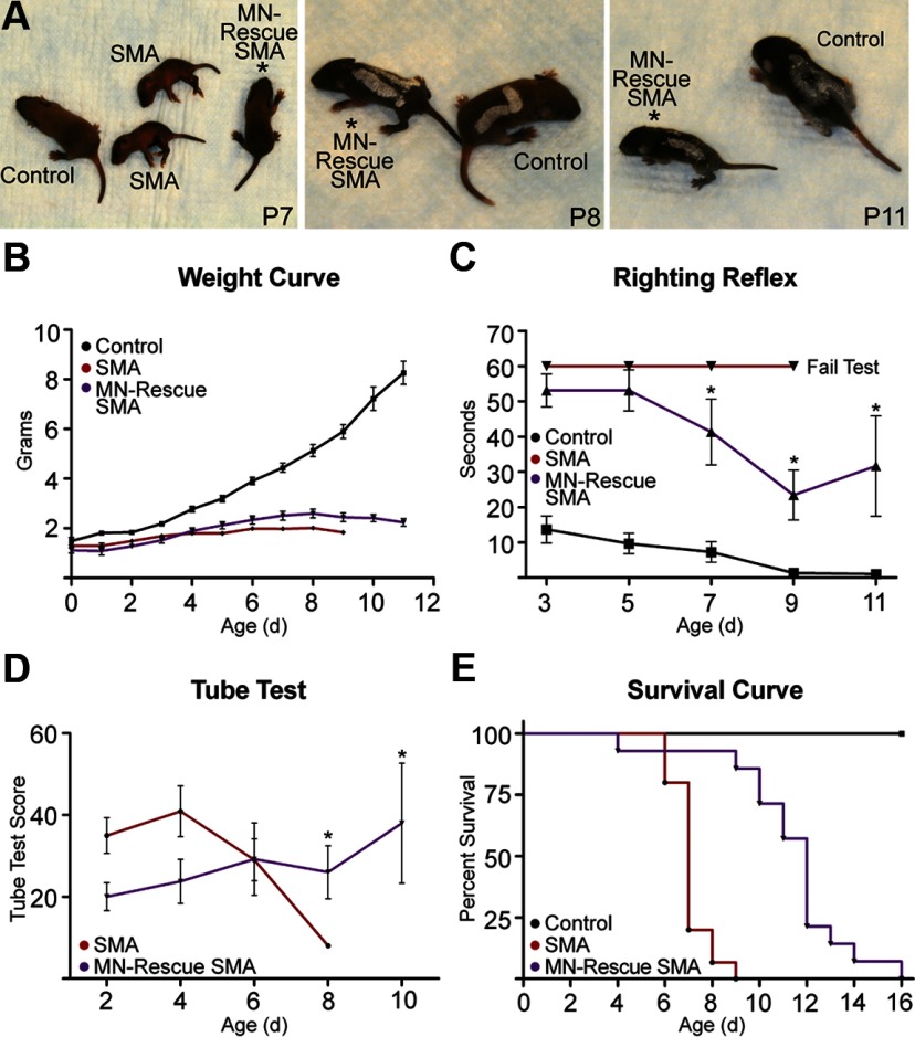
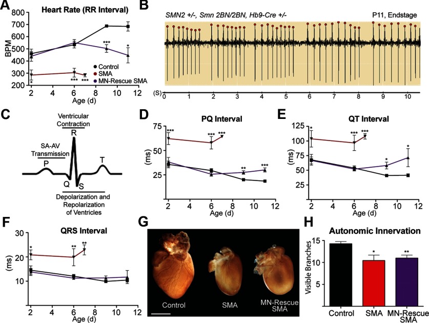
The loss of motor neurons (MNs) is a hallmark of the neuromuscular disease spinal muscular atrophy (SMA); however, it is unclear whether this phenotype autonomously originates within the MN. To address this question, we developed an inducible mouse model of severe SMA that has perinatal lethality, decreased motor function, motor unit pathology, and hyperexcitable MNs. Using an Hb9-Cre allele, we increased Smn levels autonomously within MNs and demonstrate that MN rescue significantly improves all phenotypes and pathologies commonly described in SMA mice. MN rescue also corrects hyperexcitability in SMA motor neurons and prevents sensory-motor synaptic stripping. Survival in MN-rescued SMA mice is extended by only 5 d, due in part to failed autonomic innervation of the heart. Collectively, this work demonstrates that the SMA phenotype autonomously originates in MNs and that sensory-motor synapse loss is a consequence, not a cause, of MN dysfunction.


Figures

Figure 1.
Figure 1.
Phenotypic characterization of severe inducible SMA mice. A, Western blot analysis of Smn levels. The homozygous presence of the neomycin cassette decreases Smn to 6% relative to littermate controls. This level is increased to an asymptomatic 37% if one neomycin-containing allele is replaced with a rescued allele. B, Photomicrograph. Severe inducible SMA mice were smaller and weaker in appearance than littermate controls and exhibited near complete paralysis by P5. The ruler is in centimeters. C, Kaplan–Meier survival curve. Median survival of severe inducible SMA mice is 7 d (control, n > 50; SMA, n = 15; p ≤ 0.001, log-rank test). D, SMA mice weighed significantly less than controls (P2 or later; p ≤ 0.05) and generally died after 2 consecutive days of weight loss (control, n = 16; SMA, n = 14; 2-way ANOVA, Bonferroni's post-test). E, Tube test scores in severe SMA mice were significantly diminished from the earliest detectable points (P2 or later; p ≤ 0.01; 1-way ANOVA, Bonferroni's post-test; control, n = 22; SMA, n = 15). F, SMA mice were bradycardic from the earliest point analyzed by ECG (P3; p ≤ 0.04; Student's t test; control, n = 10; SMA, n = 7). Values are shown as mean ± SEM. *p ≤ 0.05; **p ≤ 0.01; ***p ≤ 0.001.
Figure 2.
Figure 2.
Severe inducible SMA mice have presynaptic and postsynaptic defects. A–D, Micrograph images depicting control (A, C) and mutant (B, D) IC and TS NMJs, respectively. E, Representative images of NMJs that have full innervation, have partial innervation, or are denervated. F, Severe inducible (n = 7) and delta-7 SMA mice (n = 3) had a decrease in the number of fully occupied motor endplates in the ICs (p = 0.007 and p = 0.02, respectively; 2-way ANOVA, Bonferroni's post-test). G, Severe inducible and delta-7 SMA mice showed no defects in innervation of the TS. H, Examples of postsynaptic maturity. I, J, Severe inducible (n = 7) and delta-7 SMA mice (n = 3) had immature motor endplates in the ICs and protection in the TS (n = 5, control). K, L, Severe inducible (n = 7) and delta-7 SMA mice (n = 3) had NF accumulation in the ICs and exhibited resistance to disease in the TS (n = 5, control). Greater than 50 motor endplates per biological replicate were assessed for innervation, postsynaptic maturity, and presynaptic defects. Scale bars: 50 μm. Graph values are shown as mean ± SEM. *p ≤ 0.05; **p ≤ 0.01.
Figure 3.
Figure 3.
Specificity and efficiency of Hb9-Cre-driven recombination in vivo. A, A 3-primer PCR assay detects the presence or absence of the neomycin cassette in P5 MN-rescued SMA mice. Excision was observed in the spinal cord, with only minor amounts of promiscuous Cre activity in the stomach and kidney. TA, Tibialis anterior; LG, lateral gastrocnemius; Bicep, biceps brachii. A tamoxifen responsive Cre allele was used to induce severe inducible SMA pups as a positive control for excision. B, SMN2+/−, Smn2B-Neo/2B-Neo, Hb9-Cre+/−, (Cg)-Tg(CAG-Bgeo/GFP) mice show strong GFP expression in ChAT-positive cells, indicating efficient recombination specific to spinal MNs in vivo. C, Western blot. MN-rescued SMA mice showed a visible increase in Smn within the spinal cord and no increase within the ICs, kidney, or brain. D, ChAT-positive SMA motor neurons demonstrated a significant increase in the number of MNs containing nuclear gems (SMA, 17 ± 3% vs MN-rescued SMA, 51 ± 2%; p = 0.003), as well as the average number of gems per MN, confirming that Smn dosage was increased in MNs. White arrows denote gems (Table 1). Scale bars: B, 50 μm; D, 100 μm.
Figure 4.
Figure 4.
MN-rescued SMA mice have improved motor function, but minimally improved survival. A, Gross phenotype of severe inducible SMA mice is improved by Hb9-Cre. Pups are numbered using a white paint pen. B, Despite improved motor function, MN-rescued SMA mice were runted and did not show significantly improved weights (control, n > 50; SMA, n = 13; MN-rescued SMA, n = 10). C, D, MN-rescued SMA mice demonstrated a decreased level of consciousness (lethargy) early in life, resulting in poor motor function scores. This phenotype dissipates between P6 and P8, and righting reflex and tube test scores significantly improved (p ≤ 0.05 and p ≤ 0.05, respectively; 2-way ANOVA, Bonferroni's post-test; control, n ≥ 21; SMA, n = 9; MN-rescued SMA, n = 7). E, Kaplan–Meier survival curve. Despite the dramatic improvement in motor function, median survival was only increased 5 d in MN-rescued SMA mice (p ≤ 0.001, log-rank test) (control, n > 50; SMA, n = 13; MN-rescued SMA, n = 12). Values are shown as mean ± SEM. *p ≤ 0.05.
Figure 5.
Figure 5.
Hb9-Cre rescues motor unit pathology in SMA mice. A–C, At end stage, MN-rescued SMA NMJs within the ICs were indistinguishable from controls in innervation status and endplate maturity (n = 6 per genotype, >50 NMJs per mouse). D, E, NMJs within the TS of MN-rescued SMA mice showed no difference in innervation and demonstrated only a variable delay in postsynaptic maturity (n = 3 per genotype, >50 NMJs per mouse; p = 0.04; 2-way ANOVA, Bonferroni's post-test). F, G, Severe inducible SMA mice had a significant reduction in the average number of ChAT and Islet-1 double-positive MNs per ventral horn within cervical (C4–C8), thoracic (T8–T11), and lumbar (L2–L5) spinal cord (p = 0.01, p = 0.04, and p = 0.05, respectively). In contrast, MN-rescued SMA mice showed no loss in MNs in cervical and thoracic spinal segments, and had only a significant loss at the lumbar level, where Hb9-Cre expression is incomplete. Black, Control; red, SMA; purple, MN-rescue SMA. H, I, MN-rescued SMA mice did not differ from controls in average number of lumbar MMC neurons and showed a significant 25 ± 9% (p = 0.04) reduction in LMC neurons. A significant decrease in both MMC (−47 ± 12%; p = 0.05) and LMC (−29 ± 10%; p = 0.04) neurons was observed in severe inducible SMA mice (Student's t test). J, Confocal images of P5 L2–L5 motor neurons. vGlut1 labels the synapses (green), and the postsynaptic MN is marked by ChAT (red; control, n = 3; SMA, n = 3; MN-rescued SMA, n = 6; average of 44 MN per mouse). K, SMA mice had significantly fewer synapses juxtaposed to MNs per 100 μm of soma (2.74 ± 0.05, p = 0.04) relative to control littermates (3.67 ± 0.32). MN-rescued SMA mice were significantly improved relative to SMA mice (3.92 ± 0.19; p = 0.003; 1-way ANOVA, Bonferroni's post-test). Scale bars: 50 μm. Values are shown as mean ± SEM. *p ≤ 0.05; **p ≤ 0.01.
Figure 6.
Figure 6.
Whole-cell patch clamp on medial MNs. A–C, Images representative of control, SMA, and MN-rescued SMA motor neurons. MNs were filled with Texas Red during recording. D, The current command shows the protocol of hyperpolarizing and depolarizing pulses. E–G, Representative traces of fluctuations in membrane potential in control, SMA, and MN-rescued SMA motor neurons in response to voltage changes. E, F, While some postsynaptic potentials are common in the slice preparation when no blockers are present, there was an overt increase in synaptic activity in the SMA motor neurons. The frequency of postsynaptic potentials per second was quantified as significantly higher in SMA motor neurons (12.4 ± 5.1) relative to control motor neurons (3.3 ± 0.3; p = 0.05) (Table 2). G, MN-rescued SMA motor neurons had complete correction of aberrant postsynaptic potentials (2.5 ± 0.6), suggesting that this phenotype originates in MNs. Boxed insets are magnified from the boxed regions of each trace. Scale bars: C, 100 μm; D, 0.5 nA. Calibration: E–G, vertical, 20 mV; horizontal, 1 s; insets, vertical, 15 mV; horizontal, 1 s.
Figure 7.
Figure 7.
MN-rescued SMA mice have deficits in autonomic control of the heart. A, B, The heart rate of MN-rescued SMA mice (n = 7) did not differ from controls (n = 14) early in life. However, at P9, MN-rescued SMA mice became significantly bradycardic (p ≤ 0.001) and began to show hallmark signs of terminal heart block, which manifested as dropped beats and prolonged PR interval (SMA, n = 6; Student's t test). C, Schematic of the relevant components of ECG. SA, sinoatrial node; AV, atrioventricular node. D, E, MN-rescued SMA mice demonstrated deficits in aspects of ECG that are attributable to problems in sympathetic innervation of the heart, such as SA-AV signal transmission (P9–P11; p ≤ 0.01) and duration from depolarization to repolarization of the ventricles (P9–P11; p ≤ 0.05) (Student's t test). F, MN-rescued SMA mice did not differ from controls in duration of the QRS complex, suggesting that the ability of the myocardium to contract once depolarized was intact. G, H, Sympathetic innervation of the heart was visualized with tyrosine hydroxylase staining at P5. A significant loss in major visible branches was quantified in both SMA (n = 3) and MN-rescued SMA mouse hearts (n = 4) relative to controls (n = 4), suggesting that this phenotype is independent of motor unit dysfunction (Student's t test). Values are shown as mean ± SEM. Scale bar, 1 mm. *p ≤ 0.05; **p ≤ 0.01; ***p ≤ 0.001.

Similar articles

Cited by

References

    1. Araujo Ade Q, Araujo M, Swoboda KJ. Vascular perfusion abnormalities in infants with spinal muscular atrophy. J Pediatr. 2009;155:292–294. - PMC - PubMed
    1. Arber S, Han B, Mendelsohn M, Smith M, Jessell TM, Sockanathan S. Requirement for the homeobox gene Hb9 in the consolidation of motor neuron identity. Neuron. 1999;23:659–674. - PubMed
    1. Bevan AK, Hutchinson KR, Foust KD, Braun L, McGovern VL, Schmelzer L, Ward JG, Petruska JC, Lucchesi PA, Burghes AH, Kaspar BK. Early heart failure in the SMNDelta7 model of spinal muscular atrophy and correction by postnatal scAAV9-SMN delivery. Hum Mol Genet. 2010;19:3895–3905. - PMC - PubMed
    1. Boon KL, Xiao S, McWhorter ML, Donn T, Wolf-Saxon E, Bohnsack MT, Moens CB, Beattie CE. Zebrafish survival motor neuron mutants exhibit presynaptic neuromuscular junction defects. Hum Mol Genet. 2009;18:3615–3625. - PMC - PubMed
    1. Briese M, Esmaeili B, Fraboulet S, Burt EC, Christodoulou S, Towers PR, Davies KE, Sattelle DB. Deletion of smn-1, the Caenorhabditis elegans ortholog of the spinal muscular atrophy gene, results in locomotor dysfunction and reduced lifespan. Hum Mol Genet. 2009;18:97–104. - PMC - PubMed

Publication types