. 2009;9(9):7003-20.
doi: 10.3390/s90907003. Epub 2009 Sep 3.

Primary cilia: highly sophisticated biological sensors

Affiliations

Primary cilia: highly sophisticated biological sensors

Wissam A Abou Alaiwi et al. Sensors (Basel). 2009.

Abstract

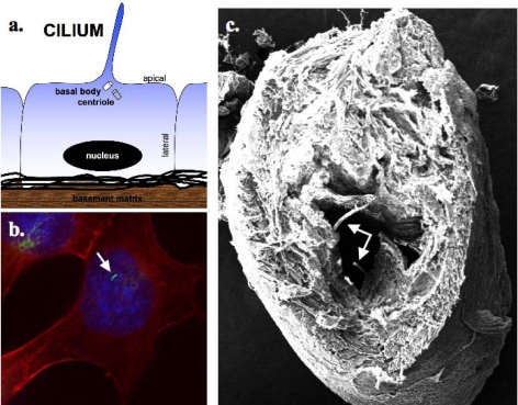
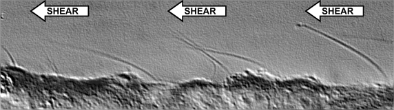
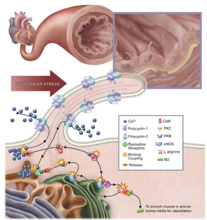
Primary cilia, thin hair-like structures protruding from the apical surface of most mammalian cells, have gained the attention of many researchers over the past decade. Primary cilia are microtubule-filled sensory organelles that are enclosed within the ciliary membrane. They originate at the cell surface from the mother centriole that becomes the mature basal body. In this review, we will discuss recent literatures on the roles of cilia as sophisticated sensory organelles. With particular emphasis on vascular endothelia and renal epithelia, the mechanosensory role of cilia in sensing fluid shear stress will be discussed. Also highlighted is the ciliary involvement in cell cycle regulation, development, cell signaling and cancer. Finally, primary cilia-related disorders will be briefly described.

Keywords: calcium; fluid shear stress; mechanosensory transduction; primary cilia.


Figures

Figure 1.
Figure 1.
Cilia are sensory organelles that project from the apical sides of cells. (a) A cartoon depicts extension of cilia from one of the centrioles, which is termed basal body. (b) Immunostaing study shows the presence of a cilium of an endothelial cell. (c) Electron micrograph further confirms the presence of cilia in the lumen of embryonic aorta in vivo. Arrows indicate the presence of cilia.
Figure 2.
Figure 2.
Cilia are sensory organelles that sense fluid-shear stress on the apical membrane of the cells. Fluid flow that produces enough drag-force on the top of the cells will bend sensory cilia. This biomechanical properties play a very important role in vestibular organs that support bodily fluid perfusion.
Figure 3.
Figure 3.
The presence of cilia in vasculature plays an important role in the biochemical production of a potent vasodilator, nitric oxide (NO). The figure depicts production of NO in an artery. Increases in blood pressure, which is translated to higher vascular shear stress, will be sensed by mechanosensory cilia. Bending or activation of the cilia involves mechanosensory polycystin-1 and polycystin-2 complex and a cascade of biochemical synthesis of NO. The cascade will further involve extracellular calcium influx (Ca2+), followed by activation of various calcium-dependent proteins, including calmodulin (CaM) and protein kinase C (PKC). Together with PKB, CaM and PKC are important downstream molecular components to activate endothelial nitric oxide synthase (eNOS).

References

    1. Satir P., Christensen S.T. Structure and function of mammalian cilia. Histochem. Cell. Biol. 2008;129:687–693. - PMC - PubMed
    1. Wheatley D.N., Wang A.M., Strugnell G.E. Expression of primary cilia in mammalian cells. Cell Biol. Int. 1996;20:73–81. - PubMed
    1. Nauli S.M., Zhou J. Polycystins and mechanosensation in renal and nodal cilia. Bioessays. 2004;26:844–856. - PubMed
    1. Rosenbaum J.L., Witman G.B. Intraflagellar transport. Nat. Rev. Mol. Cell Biol. 2002;3:813–825. - PubMed
    1. Satir P., Christensen S.T. Overview of structure and function of mammalian cilia. Annu. Rev. Physiol. 2007;69:377–400. - PubMed