. 2012 Mar 15;11(3):227-39.
doi: 10.1016/j.chom.2012.01.017.

Zinc sequestration by the neutrophil protein calprotectin enhances Salmonella growth in the inflamed gut

Affiliations

Zinc sequestration by the neutrophil protein calprotectin enhances Salmonella growth in the inflamed gut

Janet Z Liu et al. Cell Host Microbe. .

Abstract

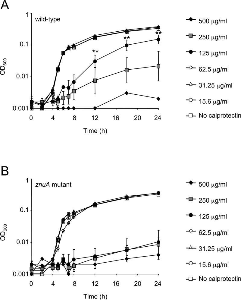
Neutrophils are innate immune cells that counter pathogens by many mechanisms, including release of antimicrobial proteins such as calprotectin to inhibit bacterial growth. Calprotectin sequesters essential micronutrient metals such as zinc, thereby limiting their availability to microbes, a process termed nutritional immunity. We find that while calprotectin is induced by neutrophils during infection with the gut pathogen Salmonella Typhimurium, calprotectin-mediated metal sequestration does not inhibit S. Typhimurium proliferation. Remarkably, S. Typhimurium overcomes calprotectin-mediated zinc chelation by expressing a high affinity zinc transporter (ZnuABC). A S. Typhimurium znuA mutant impaired for growth in the inflamed gut was rescued in the absence of calprotectin. ZnuABC was also required to promote the growth of S. Typhimurium over that of competing commensal bacteria. Thus, our findings indicate that Salmonella thrives in the inflamed gut by overcoming the zinc sequestration of calprotectin and highlight the importance of zinc acquisition in bacterial intestinal colonization.


Figures

Figure 1
Figure 1. Expression of calprotectin in the cecum of mice infected with S. Typhimurium
(A, B) S100a8 and S100a9 were detected by RT-PCR at 72 h (A) and 96 h (B) after infection with wild-type S. Typhimurium. Data are expressed as fold increase over mock-infected wild-type mice. Bars represent the geometric means ± standard error. (C, D) The concentration of calprotectin detected by ELISA (top panel) and Western Blot (bottom panel) in the fecal samples (C) and the large intestine with content (D) of mice 96 hours post infection with S. Typhimurium (n=4) or mock control (n=4). Bars represent the geometric mean ± standard deviation. ** P value ≤ 0.01. (E) Expression of the S100a8 and S100a9 mRNAs was detected in crypt colonocytes isolated from the colon of mice infected with S. Typhimurium (n=3) and expressed as fold increase over mock-infected mice (n=3). Expression of Lcn2 was detected as a positive control, and of the T cell cytokine Il-17a and the neutrophil marker Ly6g as a negative control. ND = not detected. Data represents the geometric mean ± standard error. A significant increase over mock control is indicated by * (P value ≤ 0.05) and ** (P value ≤ 0.01). (See also figure S1 and supplemental table 1).
Figure 2
Figure 2. Expression of calprotectin in mice following neutrophil depletion
Mice were injected intraperitoneally with either normal rabbit serum (NRS) or a rabbit polyclonal antibody blocking the Cxcr2 receptor (α-Cxcr2) 24 hours prior to infection with S. Typhimurium and sacrificed at 72 hours post-infection. (A) Representative dot plot (FSC=forward scatter; SSC=side scatter) of blood cells gated on leucocytes from mice treated with NRS (top) and α-Cxcr2 (bottom) (B) Representative dot plot of blood leucocytes gated on neutrophils expressing Ly6g and CD11b from mice treated with NRS (top) and α-Cxcr2 (bottom). (C, D) Cecal expression of the S100a8 and the S100a9 subunits of calprotectin was detected by Real-time RT PCR (C) and Western blot (D). (E,F) Crypt expression of the S100a8 and the S100a9 subunits of calprotectin was detected by Real-time RT PCR (E) and Western blot (F); MPO=myeloperoxidase. Data represents the geometric mean ± standard error. A significant difference in expression between NRS-treated and α-Cxcr2-treated mice is indicated by ** (P value ≤ 0.01).
Figure 3
Figure 3. Growth of S. Typhimurium in rich media supplemented with calprotectin
S. Typhimurium wild-type (A) or the znuA mutant (B) were grown in LB media supplemented with calprotectin at the indicated concentrations. Growth was determined by reading the OD600 in a microplate reader at the indicated times. Data represent the geometric mean of 4 biological replicates ± standard error. A significant difference in growth between wild-type and the znuA mutant is indicated by ** (P value ≤ 0.01). (See also figure S2 and supplemental table 2).
Figure 4
Figure 4. The ZnuABC zinc transporter promotes S. Typhimurium colonization of the inflamed cecum
(A) The concentration of zinc was measured by ICP-MS in fecal samples collected from mock-infected (n=4) or S. Typhimurium-infected (n=4) mice four days post-infection. Bars represent geometric means ± standard deviation. A significant difference is indicated by ** (P value ≤ 0.01) (B-C) Histopathology of cecal samples were collected from mice four days after infection with S. Typhimurium wild-type, the znuA mutant, or the invA spiB mutant. (B) H&E stained cecal sections from representative animals in each group. An image at lower magnification (10x) and one at higher magnification (40x) from the same section are shown. L=lumen; M=mucosa; SM=submucosa. Note marked edema in the submucosa and inflammation in mice infected with both S. Typhimurium wild-type and the znuA mutant. (C) Blinded histopathology score indicating the score of individual mice (circles), and the average score for each group (bars). The grey quadrant includes scores indicative of moderate to severe inflammation. (D) S100a8, S100a9, myeloperoxidase (MPO) and tubulin were detected by immunoblot in the cecum of mice infected with S. Typhimurium wild-type or the znuA mutant. (E) Enumeration of S. Typhimurium in the colon content (wild-type n=11, znuA mutant n=11, invA spiB mutant n=6; znuA+ZnSO4 n=9; wild-type+ZnSO4 n=6) (F) Analysis of the cecal microbiota using 16S rRNA gene qRT-PCR (wild-type n=10, znuA mutant n=9, invA spiB mutant n=6; znuA+ZnSO4 n=9). (E-F) Bars represent geometric means ± standard error. (A,E) A significant difference in comparison to wild-type infected mice is indicated by ** (P value ≤ 0.01). (F) Significant differences between groups are indicated by * (P value ≤ 0.05) and ** (P value ≤ 0.01). (See also figure S3 and supplemental table 3).
Figure 5
Figure 5. Analysis of the host response in mice infected with S. Typhimurium (wild-type + znuA mutant) or mock
(A) Histopathology of the cecum. Upper panels, H&E stained cecal sections from representative animals in each group. An image at lower magnification (10x) and one at higher magnification (40x) from the same section are shown. L=lumen; M=mucosa; SM=submucosa. Note marked edema in the submucosa and inflammation in infected mice. Lower panel, blinded histopathology scores, indicating the score of individual mice (circles), and the average score for each group (bars). (B-C) Transcript levels of Cxcl-1 (B) and Ly6g (C), were determined in wild-type mice (white bars), S100a9-/- mice (dark grey bars), and wild-type mice supplemented with zinc sulfate (light grey bars). Mice were either mock-infected or infected with S. Typhimurium as indicated. Data are expressed as fold increase over mock-infected wild-type mice. Bars represent the geometric mean of at least 4 replicates ± standard error. Significant differences in gene expression in comparison to wild-type infected C57BL/6 mice (first group) are indicated by ** (P value ≤ 0.01). (See also figure S4)
Figure 6
Figure 6. Calprotectin expression in the cecum detected by quantitative real-time PCR and Western Blot
Transcript levels of S100a8 (A) and S100a9 (B) were determined in wild-type mice (white bars), S100a9-/- mice (dark grey bars), and wild-type mice supplemented with zinc sulfate (light grey bars). Mice were either mock-infected or infected with S. Typhimurium as indicated. Bars represent the geometric mean of at least 4 replicates ± standard error. Significant differences in gene expression in comparison to wild-type infected C57BL/6 mice (first group) are indicated by * (P value ≤ 0.05) and ** (P value ≤ 0.01). (C-F) S100a8, S100a9, myeloperoxidase (MPO) and tubulin were detected by immunoblot in the cecum of mice infected with S. Typhimurium. Strain and mouse genotypes are indicated.
Figure 7
Figure 7. Resistance to calprotectin-mediated zinc sequestration provides a growth advantage to S. Typhimurium
Cecal samples were collected from mice three or four days after infection with either S. Typhimurium or mock control. Competitive index was calculated by dividing the output ratio (CFU of the wild-type /CFU of the mutant) by the input ratio (CFU of the wild-type /CFU of the mutant). (A-C) Competitive indices of S. Typhimurium strains in the colon contents of mice (n≥6/group) at four days (A) or three days (B) post infection. Strain and mouse genotypes are indicated. (C) Competitive index in cecal content of mice treated with either normal rabbit serum (NRS) or a rabbit polyclonal antibody blocking the Cxcr2 receptor (α-Cxcr2) at 72 hours post-infection. Bars represent geometric mean ± standard error. Significant differences are indicated by * (P value ≤ 0.05) and ** (P value ≤ 0.01). (See also figure S5).

Comment in

Similar articles

Cited by

References

    1. Ammendola S, Pasquali P, Pistoia C, Petrucci P, Petrarca P, Rotilio G, Battistoni A. High-affinity Zn2+ uptake system ZnuABC is required for bacterial zinc homeostasis in intracellular environments and contributes to the virulence of Salmonella enterica. Infection and immunity. 2007;75:5867–5876. - PMC - PubMed
    1. Barman M, Unold D, Shifley K, Amir E, Hung K, Bos N, Salzman N. Enteric salmonellosis disrupts the microbial ecology of the murine gastrointestinal tract. Infection and immunity. 2008;76:907–915. - PMC - PubMed
    1. Barthel M, Hapfelmeier S, Quintanilla-Martinez L, Kremer M, Rohde M, Hogardt M, Pfeffer K, Russmann H, Hardt WD. Pretreatment of mice with streptomycin provides a Salmonella enterica serovar Typhimurium colitis model that allows analysis of both pathogen and host. Infection and immunity. 2003;71:2839–2858. - PMC - PubMed
    1. Campoy S, Jara M, Busquets N, Perez De Rozas AM, Badiola I, Barbe J. Role of the high-affinity zinc uptake znuABC system in Salmonella enterica serovar Typhimurium virulence. Infection and immunity. 2002;70:4721–4725. - PMC - PubMed
    1. Conti HR, Shen F, Nayyar N, Stocum E, Sun JN, Lindemann MJ, Ho AW, Hai JH, Yu JJ, Jung JW, et al. Th17 cells and IL-17 receptor signaling are essential for mucosal host defense against oral candidiasis. The Journal of experimental medicine. 2009;206:299–311. - PMC - PubMed

Publication types

MeSH terms