. 2012 Mar 15;11(3):264-76.
doi: 10.1016/j.chom.2012.01.018.

Autophagy protein Rubicon mediates phagocytic NADPH oxidase activation in response to microbial infection or TLR stimulation

Affiliations

Autophagy protein Rubicon mediates phagocytic NADPH oxidase activation in response to microbial infection or TLR stimulation

Chul-Su Yang et al. Cell Host Microbe. .

Abstract

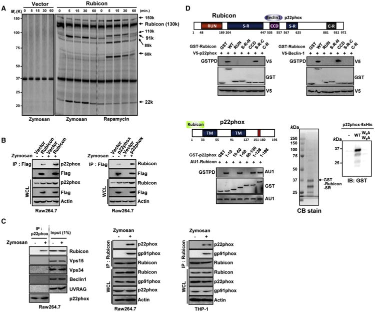
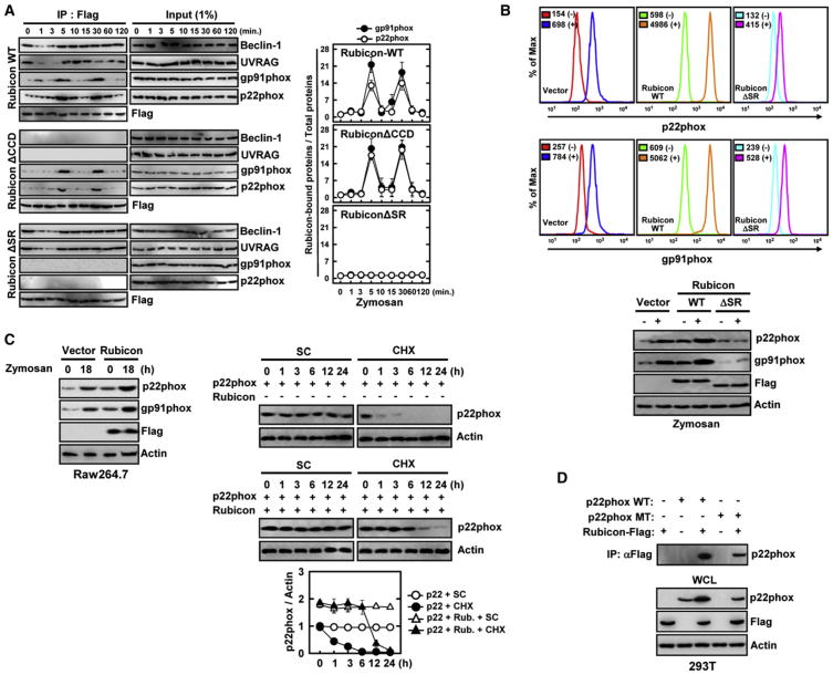
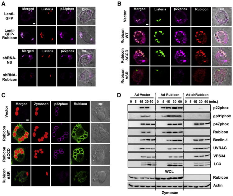
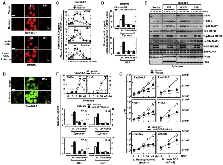
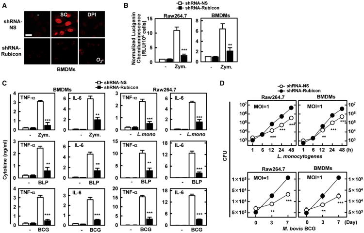
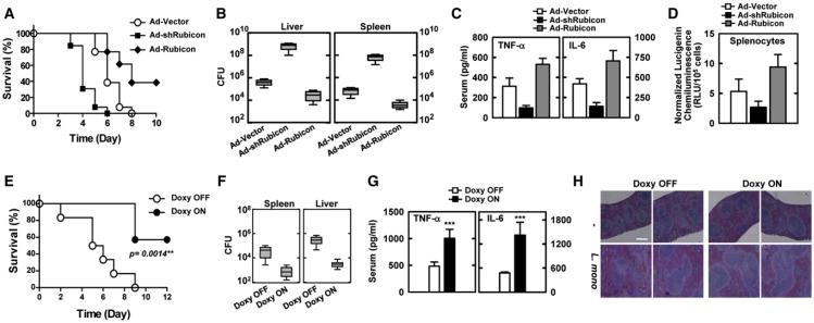
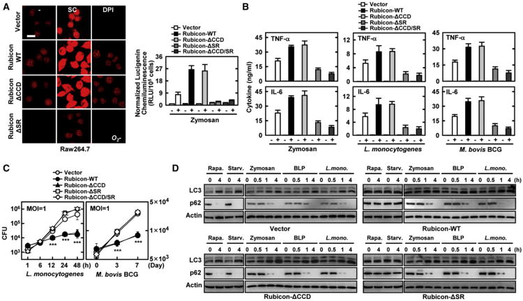
Phagocytosis and autophagy are two important and related arms of the host's first-line defense against microbial invasion. Rubicon is a RUN domain containing cysteine-rich protein that functions as part of a Beclin-1-Vps34-containing autophagy complex. We report that Rubicon is also an essential, positive regulator of the NADPH oxidase complex. Upon microbial infection or Toll-like-receptor 2 (TLR2) activation, Rubicon interacts with the p22phox subunit of the NADPH oxidase complex, facilitating its phagosomal trafficking to induce a burst of reactive oxygen species (ROS) and inflammatory cytokines. Consequently, ectopic expression or depletion of Rubicon profoundly affected ROS, inflammatory cytokine production, and subsequent antimicrobial activity. Rubicon's actions in autophagy and in the NADPH oxidase complex are functionally and genetically separable, indicating that Rubicon functions in two ancient innate immune machineries, autophagy and phagocytosis, depending on the environmental stimulus. Rubicon may thus be pivotal to generating an optimal intracellular immune response against microbial infection.


Figures

Figure 1
Figure 1. Rubicon Interaction with the p22phox-gp91phoxNADPH Oxidase Complex
(A) Differential interactions of Rubicon with the Beclin-1-containing autophagy complex and the p22phox-gp91phox complex. Radioactively labeled THP-1 cells containing vector or Flag-Rubicon were stimulated with zymosan or rapamycin for the indicated times, followed by IP with αFlag for autoradiography. (B) Rubicon interaction with p22phox. At 48 hr posttransfection with Flag-Rubicon (left) or Flag-p22phox (right), Raw264.7 cells were stimulated with or without zymosan for 30 min prior to immunoprecipitation (IP), followed by immunoblotting (IB). (C) Rubicon interaction with p22phox. Raw264.7 and THP-1 cells were stimulated with zymosan for 30 min, followed by IP with αp22phox or αRubicon and IB. (D) Binding mapping. Schematic diagram of Rubicon and p22phox. S-R, serine-rich; CCD, coiled-coil domain; C-R, cystein-rich; and TM, transmembrane (top). At 48 hr posttransfection with mammalian GST or GST-Rubicon constructs together with V5-p22phox (left) or V5-Beclin-1 (right) or GST or GST-p22phox constructs together with AU1-Rubicon (bottom left), 293T cells were used for GST pulldown, followed by IB with αV5, αGST or αAU1. (bottom right) Direct interaction between Rubicon and p22phox. Bacterially purified GST-RubiconSR-C was analyzed by Coomassie blue staining (left) or incubated with nickel beads alone, WT p22phox (N-terminal 10aa)-6xHis or its mutant W6AW9A-6xHis to evaluate direct and specific binding, followed by IB with αGST (right). See also Figure S1.
Figure 2
Figure 2. Rubicon Interaction Leads to p22phox-gp91phox Stabilization
(A) Differential Rubicon interactions. Raw264.7 cells containing vector, Flag-Rubicon WT or its mutant were stimulated with zymosan for the indicated times, followed by IP with αFlag and IB with αBeclin-1, αUVRAG, αgp91phox, αp22phox or αFlag. The right panels show the densitometry results of all three independent coIP assays. (B) (top) Rubicon-mediated increases of p22phox and gp91phox levels. THP-1 cells containing vector, Rubicon WTor ΔSR were stimulated with (−) or without zymosan (+) for 18 hr and used for flow cytometry analysis to detect intracellular p22phox or surface expressing gp91phox. (bottom) IB with αp22phox, αgp91phox, αflag and αActin. (C) Rubicon-mediated increases of p22phox and gp91phox stability. (left) Raw264.7 cells containing vector or Flag-Rubicon were stimulated with zymosan for 18 hr, followed by IB with αp22phox, αgp91phox, αflag and αActin. (right) At 24 hr post-transfection with p22phox and/or Flag-Rubicon, 293T cells were treated with solvent control (SC) or cyclohexamide (CHX, 1 μg/ml) for indicated times and cell lysates were used for IB with αp22phox and αActin. The bottom panel shows the ratio of p22phox/Actin during CHX time course treatment. (D) Analysis of p22phoxY121H mutant expression upon Rubicon expression. At 48 hr post-transfection with Flag-Rubicon and/or p22phox WTor Y121H mutant, 293T cell lysates were used for IP with αFlag and IB with αp22phox. WCLs were used for IB with αp22phox, αFlag, or αActin. See also Figure S2.
Figure 3
Figure 3. Rubicon Affects p22phoxPhagosome Recruitment and Phagocytosis
(A–C) (A) Expression or depletion of Rubicon affects the colocalization of p22phox with L. monocytogenes-containing phagosomes. At 48 hr postinfection with lenti-GFP or lenti-GFP-Rubicon (top) at MOI =100 (Figure S3A) or with lenti-shRNA-NS or lenti-shRNA-Rubicon (bottom) at MOI =50 (Figure S3B), Raw264.7 cells were infected with HK-TRITC-labeled L. monocytogenes (MOI = 1) for 30 min, followed by confocal microscopy with αp22phox. Bar, 2 μm. Rubicon enhances the colocalization of p22phox with L. monocytogenes (B) or zymosan particles-containing phagosomes (C) in a p22phox-binding-dependent manner. Raw264.7 cells containing vector, Rubicon WT, ΔCCD or ΔSR were infected with HK-GFP-L. monocytogenes (B) or Texas red-labeled opsonized-zymosan particles (C) for 30 min, followed confocal microscopy with αp22phox or αFlag. Bar, 2 μm. (D) Rubicon enhances phagocytosis in a p22phox-binding-dependent manner. At 48 hr postinfection with recombinant Ad-Vector (MOI = 200), Ad-Rubicon (MOI = 100), or Ad-shRubicon (MOI = 200) virus, Raw264.7 cells were stimulated with zymosan-coated particles for indicated times, followed by lysis and sucrose-gradient ultracentrifugation to isolate the bead-containing phagosomal fractions. Phagosomal fractions were subjected to IB with αBeclin-1, αUVRAG, αgp91phox, αp22phox, αp47phox, αVPS34, αLC3 or αRubicon. WCL were used for IB with αRubicon or αActin. See also Figure S3.
Figure 4
Figure 4. Rubicon Activates NADPH Oxidase Activity in a p22phox-Binding-Dependent Manner
(A and B) Rubicon activates ROS production. Raw264.7 cells or BMDMs were incubated with 20 μM DPI (A) and 20 mM NAC (B) to detect O2 and H2O2 production, respectively, with or without zymosan for 30 min. Live cells were washed with serum-free medium and imaged using a confocal microscope. Bar, 10 μm. (C) Luminometry of NADPH oxidase activity of Raw264.7 cells containing vector or Rubicon after treatment with zymosan or BLP, or infected with L. monocytogenes (MOI = 1). (D) Rubicon enhances NADPH oxidase activity. BMDMs infected with lenti-GFP or lenti-GFP-Rubicon was analyzed for NADPH oxidase activity upon zymosan or BLP treatment. (E) Rubicon expression enhances downstream signaling. Raw264.7 cells containing vector, Rubicon or its mutants were stimulated with zymosan for the indicated times and then subjected to IB with the phosphorylated and total forms of p38, p42/p44 MAPK, JNK, IκB-α, or Actin. (F) Increase of cytokine production by Rubicon. (top) Raw264.7 cells containing vector or Rubicon were stimulated with zymosan for the indicated times and the supernatants were analyzed for cytokine ELISA. (middle and bottom) BMDMs infected with lenti-GFP or lenti-GFP-Rubicon were stimulated with zymosan or BLP for 18 hr and the supernatants were subjected to cytokine ELISA. (G) Rubicon enhances bacterial killing activity. Raw246.7, THP-1 or BMDMs containing vector or Rubicon were infected with L. monocytogenes (left) or M. bovis BCG (right) at a MOI = 1 for the indicated times and then lysed to determine intracellular bacterial loads. CFU, colony-forming units. *p < 0.05; **p < 0.01; ***p < 0.001 compared with the vector control. All data above are the mean ± SD of values from three experiments. See also Figure S4.
Figure 5
Figure 5. Rubicon's Effect on ROS and Inflammatory Cytokine Production
(A and B) Depletion of Rubicon gene expression leads to the reduction of ROS production (A) and NADPH oxidase activity (B). At 48 hr post-infection with lentivirus-shRNA-NS or lentivirus-shRNA-Rubicon (MOI = 50), Raw264.7 cells or BMDMs were analyzed for O2 production upon zymosan treatment for 30 min with or without pretreatment with DPI (A) or for NADPH oxidase activity (B) as described in Figure 4. Bar, 10 μm. (C) Reduction of cytokine production by Rubicon gene depletion. At 48 hr postinfection with lentivirus-shRNA-NS or lentivirus-shRNA-Rubicon, Raw264.7 cells or BMDMs were stimulated with zymosan, BLP, L. monocytogenes or M. bovis BCG for 18 hr, and the supernatants were analyzed for cytokine production using ELISA. (D) Rubicon gene depletion reduces bacterial killing activity. At 48 hr postinfection with lentivirus-shRNA-NS or lentivirus-shRNA-Rubicon, Raw264.7 cells or BMDMs were infected with L. monocytogenes (top) or M. bovis BCG (bottom) at a MOI = 1 for indicated times and then lysed to determine intracellular bacterial loads. **p < 0.01; ***p < 0.001 compared with the lentivirus-shRNA-NS culture. All data above are the mean ± SD of values from three experiments. See also Figure S5.
Figure 6
Figure 6. Alteration of Rubicon Gene Expression Affects Mice Mortality after L. monocytogenes Infection
(A–D) At 48 hr post-injection with Ad-vector (1 × 10¹³ pfu/kg), Ad-shRubicon (1 × 10¹² pfu/kg),or Ad-Rubicon (1 × 10¹³ pfu/kg) twice intravenously via the tail vein, mice were infected with L. monocytogenes (1 × 10⁷ CFU/mouse) and mortality was measured for n = 23 mice per group (A). Bacterial loads of infected mice (n =5 per group) in spleen and liver (B), serum cytokine levels (C) or splenocyte ROS levels (D) were determined at 5 days post-infection with L. monocytogenes (1 × 10⁶ CFU/mouse). (E–H) Doxycycline-inducible, macrophage-specific expression of Rubicon in SRA-rtTA mice. After 7 days of doxycycline treatment, mice were infected with L. monocytogenes (1 × 10⁷ CFU per mouse) and mortality was measured for n = 6 mice per group (E). L. monocytogenes loads of infected mice (n=6 per group) in liver and spleen (F). Serum cytokine levels (G) or H&E staining (I) were determined at 6 days post-infection with L. monocytogenes (1 × 10⁶ CFU/mouse). CFU, colony-forming units. ***p < 0.001 compared with the doxycycline-off conditions. The data are the mean ± SD of values from three experiments. Bar, 200 μm. See also Figure S6.
Figure 7
Figure 7. ROS-Mediated Bacterial Killing Activity is p22phox-Binding-Dependent
(A) Rubicon enhances ROS production in a p22phox-binding-dependent manner. Raw246.7 cells expressing vector, Rubicon WT, or mutants were used for O2 production (left) or for NADPH oxidase activity (right) with or without zymosan for 30 min, as described in Figure 4. Bar, 10 μm. (B) Rubicon increases cytokine production. Raw264.7 cells containing vector, Rubicon WT or its mutant were stimulated with zymosan, L. monocytogenes, or M. bovis BCG for 18 h and the supernatants were analyzed for cytokine ELISA. (C) Rubicon enhances bacterial killing activity. Raw246.7 containing vector, Rubicon WT or its mutant were infected withL. monocytogenes(left) or M. bovis BCG (right) at a MOI = 1 for the indicated times and then lysed; intracellular bacteria were plated to determine CFU. The data are the mean ± SD of values from three experiments. ***p < 0.001 compared with the vector control. (D) Rubicon plays distinctive roles in conventional and TLR-signaling-mediated autophagy. Raw246.7 cells expressing vector, Rubicon WT, ΔCCD, or ΔSR mutant were treated with rapamycin, starvation, zymosan, BLP or L. monocytogenes for indicated times and their cell lysates were used forIBwith αLC3, αp62, or αactin. See also Figure S7.

Comment in

References

    1. Bedard K, Krause KH. The NOX family of ROS-generating NADPH oxidases: physiology and pathophysiology. Physiol Rev. 2007;87:245–313. - PubMed
    1. Deretic V. Autophagy in immunity and cell-autonomous defense against intracellular microbes. Immunol Rev. 2011;240:92–104. - PMC - PubMed
    1. Deretic V, Klionsky DJ. How cells clean house. Sci Am. 2008;298:74–81. - PubMed
    1. Deretic V, Levine B. Autophagy, immunity, and microbial adaptations. Cell Host Microbe. 2009;5:527–549. - PMC - PubMed
    1. Fang FC. Antimicrobial reactive oxygen and nitrogen species: concepts and controversies. Nat Rev Microbiol. 2004;2:820–832. - PubMed

Publication types

MeSH terms