. 2012 Mar 15;11(3):298-305.
doi: 10.1016/j.chom.2012.01.014.

Pancreatic acinar cell-specific autophagy disruption reduces coxsackievirus replication and pathogenesis in vivo

Affiliations

Pancreatic acinar cell-specific autophagy disruption reduces coxsackievirus replication and pathogenesis in vivo

Mehrdad Alirezaei et al. Cell Host Microbe. .

Abstract

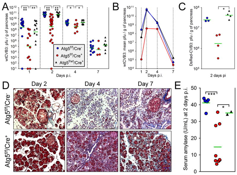
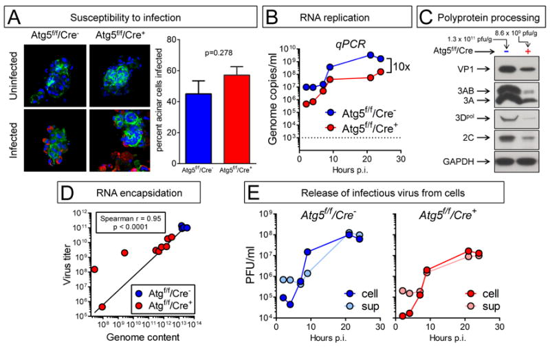
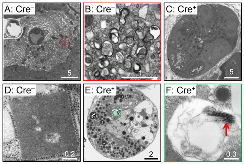
Autophagy protects against many infections by inducing the lysosomal-mediated degradation of invading pathogens. However, previous in vitro studies suggest that some enteroviruses not only evade these protective effects but also exploit autophagy to facilitate their replication. We generated Atg5(f/f)/Cre(+) mice, in which the essential autophagy gene Atg5 is specifically deleted in pancreatic acinar cells, and show that coxsackievirus B3 (CVB3) requires autophagy for optimal infection and pathogenesis. Compared to Cre(-) littermates, Atg5(f/f)/Cre(+) mice had an ∼2,000-fold lower CVB3 titer in the pancreas, and pancreatic pathology was greatly diminished. Both in vivo and in vitro, Atg5(f/f)/Cre(+) acinar cells had reduced intracellular viral RNA and proteins. Furthermore, intracellular structural elements induced upon CVB3 infection, such as compound membrane vesicles and highly geometric paracrystalline arrays, which may represent viral replication platforms, were infrequently observed in infected Atg5(f/f)/Cre(+) cells. Thus, CVB3-induced subversion of autophagy not only benefits the virus but also exacerbates pancreatic pathology.


Conflict of interest statement

The authors have no conflicting financial interests.

Figures

Figure 1
Figure 1. The pancreata of Atg5f/f/Cre+ mice are depleted of functional Atg5 protein, but the mice show little sign of constitutive pancreatic dysfunction
Generation of transgenic mice, and characterization of uninfected animals. (A). Diagram of the three Atg5 genotypes relevant to this study, “floxed”, wt, and deleted (Atg5f × Cre). Black arrowhead = lox p, black rectangle = exon of Atg5. The locations of the four primers (A-D) are shown; for sequences, see Experimental Procedures (online). The results of PCR of the three indicated genotypes are shown. All other panels in the figure show data from only two of the mouse lines: Atg5f/f/Cre+ ± Atg5f/f/Cre mice. (B). Western blots of representative pancreata (two per mouse line). The right panel shows a graph of the average OD, normalized against GAPDH signal (mean + SE). The body weights (C) and blood glucose levels (D) of age- and gender-matched mice are shown (green lines indicate means). (E). Enzyme contents of pancreata from three mice of each line; L = liver (negative control). The related graph shows the average OD in each mouse line (mean + SE). Histological sections, stained with Masson’s trichrome, are shown in panel F, and G shows an electron micrograph of pancreas from an Atg5f/f/Cre+ mouse; vacuoles are indicated by red arrows, and a severely abnormal acinar cell is encircled by a dashed red line.
Figure 2
Figure 2. In the absence of an intact autophagy pathway, pancreatic titers of infectious CVB3 are dramatically reduced early in infection, and pancreatic pathology is partially mitigated
The key in panel A, showing the three lines of mice, applies also to panels B, C & E. (A & B). Mice were infected with wtCVB3 (104 pfu i.p.) and, at days 1, 2, 4 and 7 p.i. the pancreata were harvested and virus titers of pancreatic homogenates were determined by plaque assay. In each group, the total number of mice used was: Atg5f/f/Cre, 52; Atg5f/f/Cre+, 50; Atg5f/wt/Cre+, 47. (A). Horizontal green bars indicate the geometric means, which are re-plotted in (B) to more clearly present the viral replication kinetics in each group. (C). Mice were infected with a recombinant CVB3, DsRed-CVB3 (107 pfu i.p.), and pancreatic titers were measured at 2 days p.i. (D). At the indicated times p.i., histological sections were prepared from pancreata of Atg5f/f/Cre or Atg5f/f/Cre+ mice and stained with Masson’s trichrome. (E). Serum amylase levels were measured at 2 days p.i. in the indicated mouse strains. Prior to infection, the average level was ~6 U/mL.
Figure 3
Figure 3. No single feature of the CVB3 lifecycle can be identified as being acutely Atg5-dependent in acinar cells, and responsible for the reduced in vivo replication and pathogenesis
Panels A, B, E: Acinar cells were isolated from the pancreata of uninfected Atg5f/f/Cre and Atg5f/f/Cre+ mouse lines, and were infected in vitro with wtCVB3 (MOI ~100). Panels C & D: Atg5f/f/Cre+ and Atg5f/f/Cre mice were infected with CVB3, and were sacrificed two days later. The pancreata were recovered and homogenized. (A). F-actin was detected using Alexa Fluor 488-phalloidin (green), and CVB3 VP1 using a specific antibody (red) The graph shows the proportion of each cell type that was infected (mean + SE). (B). Viral RNA content in isolated acinar cells was followed over a 24-hour period by qPCR. The dashed line shows the lower limit of detection; uninfected cells scored below this line. (C). Western immunoblot analysis of viral protein content at 2 days p.i. in the pancreata of two representative mice. The virus titer in each mouse is shown above each column. (D). Infectious virus titers in pancreata were measured by plaque assay, and genomic CVB3 RNA content was determined by qPCR. Each symbol represents a single mouse, and a Spearman correlation line for the relationship between virus titer (y axis) and viral genomic RNA content (x axis) is shown. (E) One-step growth curve in both cell types (Cre and Cre+); cell-associated (cell) and extracellular (sup) infectious virus titers were measured.
Figure 4
Figure 4. Electron microscopy of CVB3-infected Atg5f/f/Cre and Atg5f/f/Cre+ acinar cells
A-C: Pancreata were studied at 2 days p.i. A-B, Atg5f/f/Cre mice; C, Atg5f/f/Cre+ animal. (A) Infected acinar cell in a Cre pancreas, showing extensive RER remodelling. The region enclosed in a red dashed square is magnified in B, and contains compound membrane vesicles. (C) Infected cell in a Cre+ pancreas; vacuolization is present, but extensive RER remodelling, and CMVs, are absent. D-F: Acinar cells were isolated from Atg5f/f/Cre mice (D) or from Atg5f/f/Cre+ animals (E,F). The cells were infected with CVB3 (MOI = ~100) and, 18 hours later, the cells were evaluated by EM. Paracrystalline arrays, adjacent to ribosomes, and comprising subunits of ~8 nm diameter, were present in Cre cells (D) but were less frequently observed in Cre+ cells, even though the latter cells were infected, as indicated by the vacuolization that is visible at low magnification (E), and by the presence of virions, revealed at high magnification (F, red arrow). Scale bars are show for each image, and the number immediately above each bar indicates its size (microns).

References

    1. Bienz K, Egger D, Pfister T, Troxler M. Structural and functional characterization of the poliovirus replication complex. J Virol. 1992;66:2740–2747. - PMC - PubMed
    1. Brabec-Zaruba M, Berka U, Blaas D, Fuchs R. Induction of autophagy does not affect human rhinovirus type 2 production. J Virol. 2007;81:10815–10817. - PMC - PubMed
    1. Chen J, Noueiry A, Ahlquist P. An alternate pathway for recruiting template RNA to the brome mosaic virus RNA replication complex. J Virol. 2003;77:2568–2577. - PMC - PubMed
    1. Deretic V, Levine B. Autophagy, immunity, and microbial adaptations. Cell Host Microbe. 2009;5:527–549. - PMC - PubMed
    1. Gebhard JR, Perry CM, Harkins S, Lane T, Mena I, Asensio VC, Campbell IL, Whitton JL. Coxsackievirus B3-induced myocarditis: perforin exacerbates disease, but plays no detectable role in virus clearance. Am J Pathol. 1998;153:417–428. - PMC - PubMed

Publication types

Substances

LinkOut - more resources