

Review
doi: 10.1067/j.cpsurg.2011.12.002.
Unexpected gynecologic findings during abdominal surgery
Affiliations
- PMID: 22424211
- PMCID: PMC3313456
- DOI: 10.1067/j.cpsurg.2011.12.002
Item in Clipboard
Review
Unexpected gynecologic findings during abdominal surgery
Curr Probl Surg.
2012 Apr.
No abstract available
Figures
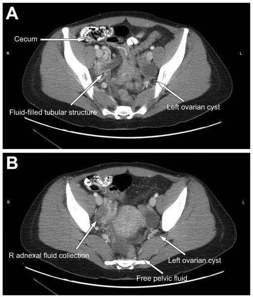
(A) A CT scan of a 24-year-old female with fever, nausea, vomiting, and abdominal pain. An enlarged, fluid-filled tubular structure is noted in the pelvis, remote from the cecum. (B) More distal cut shows a right adnexal fluid collection and a left ovarian cyst (arrows).
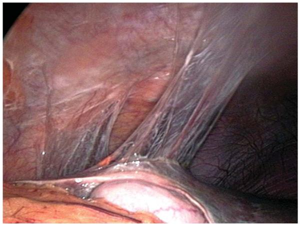
Laparoscopic view of perihepatic adhesions consistent with Fitz-Hugh-Curtis Syndrome. (Source: http://commons.wikimedia.org/wiki/File:Perihepatic_adhesions.jpg .) (Color version of figure is available online.)
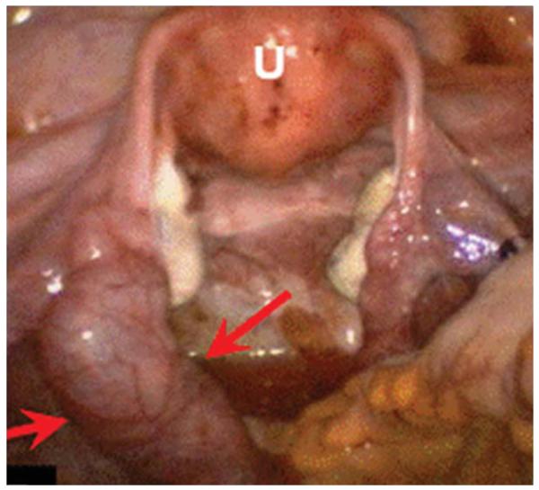
Laparoscopic view of pelvic inflammatory disease with pyosalpinx (arrows). U, uterus. (Reprinted with permission from Chandra S. ROLE OF LAPAROSCOPY IN THE MANAGEMENT OF PELVIC INFLAMATION DISEASE/TUBO-OVARIAN ABSCESS COMPARE TO OTHER MODALITIES. Available at: http://www.laparoscopyhospital.com/role-of-laparoscopy-in-the-management-of-pelvic-inflamation-disease-tubo-ovarian-abscess-compare-to-other-modalites.html .) (Color version of figure is available online.)
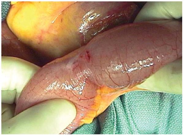
Endometrial implants on the serosa of the terminal ileum. (Reprinted with permission from Chaer R, Sam A 2nd, Teresi M, Cintron J. Endometriosis-induced acute small and large bowel obstruction: rare clinical entities. N Z Med J 2005;118:U1521. (Color version of figure is available online.)
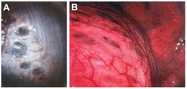
A: Thoracoscopic view of endometriosis of the diaphragm in a patient with catamenial pneumothorax. B: Thoracoscopic view of endometriosis of the diaphragm in the patient’s twin sister, also with catamenial pneumothorax. (Reprinted with permission from http://catamenialpneumothorax.com/id15.htm .) (Color version of figure is available online.)
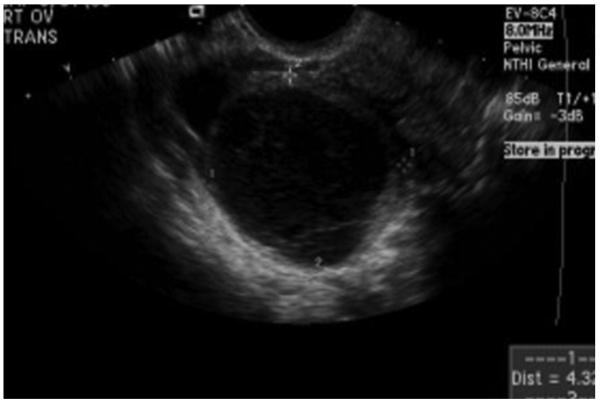
Ultrasound demonstrating a hemorrhagic ovarian cyst. Internal septations and a reticular pattern appear within the cyst as the blood begins to clot.
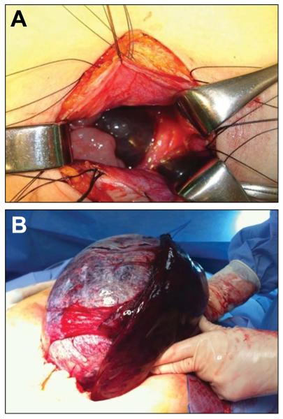
A: Intraoperative view of adnexal torsion. B: Intraoperative picture of a large, torsed ovary. (Color version of figure is available online.)
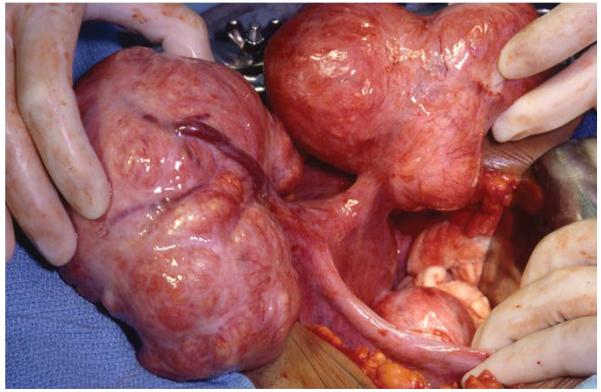
Intraoperative picture of uterine fibroids. (Color version of figure is available online.)
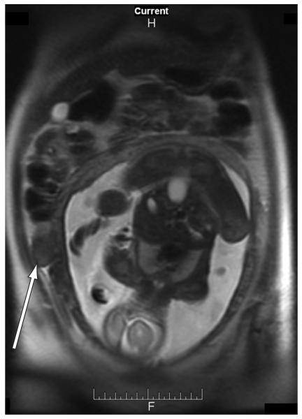
T2-weighted coronal MRI demonstrating right lower quadrant fibroid (arrow) mimicking appendicitis during pregnancy.
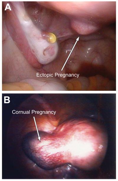
A: Laparoscopic view of cervical ectopic pregnancy. B: Laparoscopic view of cornual ectopic pregnancy. (Color version of figure is available online.)
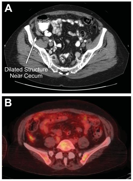
A: CT scan from a female patient with abdominal pain and a dilated structure near the cecum. The patient had previously had an appendectomy and was ultimately diagnosed with metastatic ovarian carcinoma. B: PET scan from the same patient, showing increased activity in the ovarian mass, liver, and omentum. (Color version of figure is available online.)
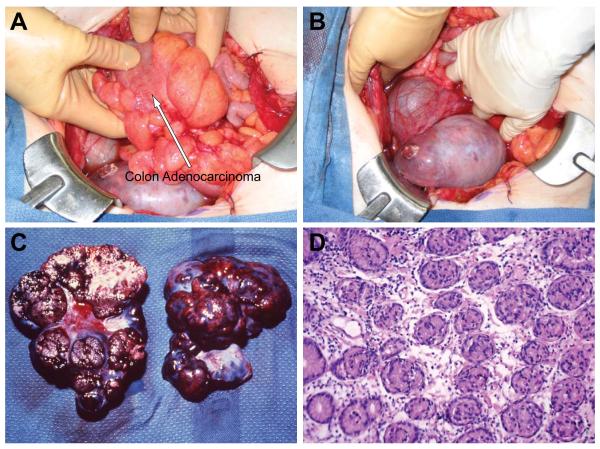
A: Intraoperative view of primary adenocarcinoma of the colon (arrow). B: Intraoperative view of ovarian metastases from adenocarcinoma of the colon in the same patient. Bilateral oophorectomy was performed. C: Bilobed ovarian mass after oophorectomy for ovarian metastases from primary colon adenocarcinoma. D: Microscopic view of colon adenocarcinoma metastatic to the ovary. (Color version of figure is available online.)
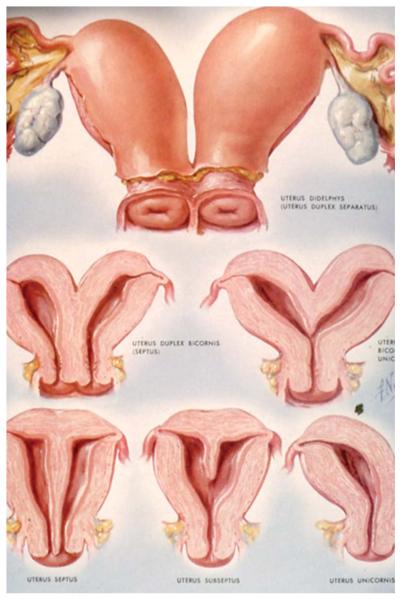
Spectrum of unification defects of the uterus. Arcuate uterus presents with small midline septum and a minimal fundal cavity indentation. A septate uterus is the most common Müllerian duct anomaly and develops when resorption of the intervening septum is incomplete. Bicornuate uterus develops due to failure of complete fusion of the Müllerian ducts; two endometrial cavities and a single cervix and vagina result. (Color version of figure is available online.)
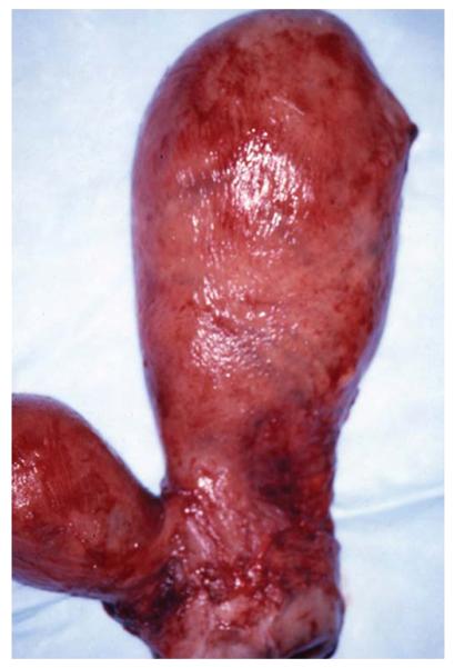
Bicornuate uterus of a 13-year-old who presented with acute right lower quadrant abdominal pain at the onset of her menses due to an undrained uterine horn. (Color version of figure is available online.)
References
-
- Karam AR, Birjawi GA, Sidani CA, et al. Alternative diagnoses of acute appendicitis on helical CT with intravenous and rectal contrast. Clin Imaging. 2007 Mar-Apr;31(2):77–86. - PubMed
-
- Vandermeer FQ, Wong-You-Cheong JJ. Imaging of acute pelvic pain. Clin Obstet Gynecol. 2009 Mar;52(1):2–20. - PubMed
-
- Silen W. Cope’s Early Diagnosis of the Acute Abdomen. 21 ed Oxford University Press; Oxford: 2005.
-
- Eshed I, Halshtok O, Erlich Z, et al. Differentiation between right tubo-ovarian abscess and appendicitis using CT-A diagnostic challenge. Clin Radiol. 2011 Jun 29; - PubMed
-
- Lewis FR, Holcroft JW, Boey J, et al. Appendicitis. A critical review of diagnosis and treatment in 1,000 cases. Arch Surg. 1975 May;110(5):677–684. - PubMed
Publication types
MeSH terms
Grants and funding
LinkOut - more resources
Full Text Sources
Medical
