Review
. 2012 Mar 16;148(6):1132-44.
doi: 10.1016/j.cell.2012.02.032.

Cellular metabolism and disease: what do metabolic outliers teach us?

Affiliations
Review

Cellular metabolism and disease: what do metabolic outliers teach us?

Ralph J DeBerardinis et al. Cell. .

Abstract

An understanding of metabolic pathways based solely on biochemistry textbooks would underestimate the pervasive role of metabolism in essentially every aspect of biology. It is evident from recent work that many human diseases involve abnormal metabolic states--often genetically programmed--that perturb normal physiology and lead to severe tissue dysfunction. Understanding these metabolic outliers is now a crucial frontier in disease-oriented research. This Review discusses the broad impact of metabolism in cellular function and how modern concepts of metabolism can inform our understanding of common diseases like cancer and also considers the prospects of developing new metabolic approaches to disease treatment.


Figures

Figure 1
Figure 1. An overview of intermediary metabolism
A simplified view of core metabolism, focusing on the use of major nutrients (glucose, amino acids and fatty acids) to produce or store energy, and to grow.
Figure 2
Figure 2. Metabolism helps to implement cell growth programs
In mammals, cell growth and proliferation are controlled by extracellular factors. These ligands bind to cell surface receptors and initiate signal transduction cascades, stimulating numerous cellular activities to enable growth and replicative division. Proper control of metabolism is required for these effects. One of the proximal effects of growth factor signaling is to increase surface expression of transporters for glucose and other nutrients, which provide energy and metabolic precursors to produce macromolecules. Catabolism of these nutrients (heavy arrows) produces carbon dioxide and energy. If nutrients are present in excess, so that flux through these basic catabolic pathways is satisfied, other pathways stemming from core metabolism are induced to propagate growth signals. Hexosamine biosynthesis reinforces growth signals by enabling cells to maintain surface expression of growth factor receptors and nutrient transporters. Acetyl-CoA generated by acetyl-CoA synthetases (ACS) and ATP-citrate lyase (ACL) provides substrate for the synthesis of lipids and other macromolecules, and for acetylation reactions to regulate gene expression and enzyme function. The favorable energy state during growth factor signaling also suppresses AMPK, thereby permitting cells to engage in energy-consuming biosynthetic pathways and to progress through the cell cycle.
Figure 3
Figure 3. Cancer cell metabolism
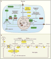
(A) Cancer cells rely primarily on glucose and glutamine to supply intermediary metabolism. Several metabolite pools fed by these nutrients and thought to be essential for tumor cell growth are highlighted in yellow. Uptake and catabolism of glucose and glutamine is regulated by oncogenic signaling. Suspected metabolic tumor suppressors (red) and oncogenes (green) control the abundance of a handful of key metabolites (bold) that regulate additional signaling functions as described in the text. These signaling activities likely contribute to malignant transformation or the propagation of growth signals within transformed cells. Thus, in addition to their traditional roles in metabolism, FH, SDH and the 2-HG dehydrogenases serve to suppress levels of pro-oncogenic metabolites. 2SC, S-(2-succinyl)-cysteine. (B) Detailed view of selected metabolites and enzymes discussed in the text. Abbreviations: Aco, aconitase; IDH, isocitrate dehydrogenase; αKGDH, α-ketoglutarate dehydrogenase; SCS, succinyl-CoA synthetase; SDH, succinate dehydrogenase; FH, fumarate hydratase; mut IDH1/IDH2, mutant isocitrate dehydrogenase 1 or 2; (D)-2HG DH; (D)-2-hydroxyglutaric acid dehydrogenase.
Figure 4
Figure 4. Metabolic flux analysis
(A) Analysis of metabolism is similar in principle to the analysis of traffic patterns, with many of the same uncertainties. The high “flux” on a four-lane highway leads to a low density of cars, all of which travel unimpeded southward. Upon exiting the highway, drivers experience reduced flux because of the traffic light, akin to mutation or under-expression of a metabolic enzyme. This causes an increased density of cars north of the light. Flux downstream of the block is unimpeded. Note that red cars on the two-lane road also merge with black cars, leading to a reduced fraction of red cars downstream of the intersection. The sum effect of these factors on overall flux is demonstrated by counting the cars that pass the checkered flags. On the highway, 1000 cars, all red, pass in one hour. On the two-lane road, only 200 cars pass, and only half are red. (B) Simple schematic of metabolic flux analysis. Glucose labeled with 13C at positions 1 and 6 (red asterisks) is given via injection or oral administration to a subject, which metabolizes it. After a period of time, tissue or body fluids are sampled to determine the abundance of various metabolites, the fraction of the metabolite that contains 13C, and the position(s) of 13C within the molecule. Data are acquired using mass spectrometry or NMR spectroscopy. Mathematical models are then applied to translate the data into metabolic flux. In this example, labeling of lactate and acetyl-CoA are examined. The pathways producing these two metabolites diverge at pyruvate. LDH, a highly active enzyme, rapidly converts pyruvate to lactate, resulting in a very high enrichment in the lactate pool in a short time. Meanwhile, two factors conspire to reduce enrichment in acetyl-CoA. First, this pathway involves PDH, a highly regulated and less active enzyme. Second, entry of carbon from unlabeled nutrients contributes to the acetyl-CoA pool, reducing the fraction of acetyl-CoA molecules containing 13C from glucose.

References

    1. Adam J, Hatipoglu E, O’Flaherty L, Ternette N, Sahgal N, Lockstone H, Baban D, Nye E, Stamp GW, Wolhuter K, et al. Renal Cyst Formation in Fh1-Deficient Mice Is Independent of the Hif/Phd Pathway: Roles for Fumarate in KEAP1 Succination and Nrf2 Signaling. Cancer Cell. 2011;20:524–537. - PMC - PubMed
    1. Alderson NL, Wang Y, Blatnik M, Frizzell N, Walla MD, Lyons TJ, Alt N, Carson JA, Nagai R, Thorpe SR, et al. S-(2-Succinyl)cysteine: a novel chemical modification of tissue proteins by a Krebs cycle intermediate. Arch Biochem Biophys. 2006;450:1–8. - PubMed
    1. Alfardan J, Mohsen AW, Copeland S, Ellison J, Keppen-Davis L, Rohrbach M, Powell BR, Gillis J, Matern D, Kant J, et al. Characterization of new ACADSB gene sequence mutations and clinical implications in patients with 2-methylbutyrylglycinuria identified by newborn screening. Mol Genet Metab. 2010;100:333–338. - PMC - PubMed
    1. Baysal BE. Clinical and molecular progress in hereditary paraganglioma. J Med Genet. 2008;45:689–694. - PubMed
    1. Baysal BE, Ferrell RE, Willett-Brozick JE, Lawrence EC, Myssiorek D, Bosch A, van der Mey A, Taschner PE, Rubinstein WS, Myers EN, et al. Mutations in SDHD, a mitochondrial complex II gene, in hereditary paraganglioma. Science. 2000;287:848–851. - PubMed

Publication types

LinkOut - more resources