Review
. 2012 Mar 16;148(6):1145-59.
doi: 10.1016/j.cell.2012.02.035.

Mitochondria: in sickness and in health

Affiliations
Review

Mitochondria: in sickness and in health

Jodi Nunnari et al. Cell. .

Abstract

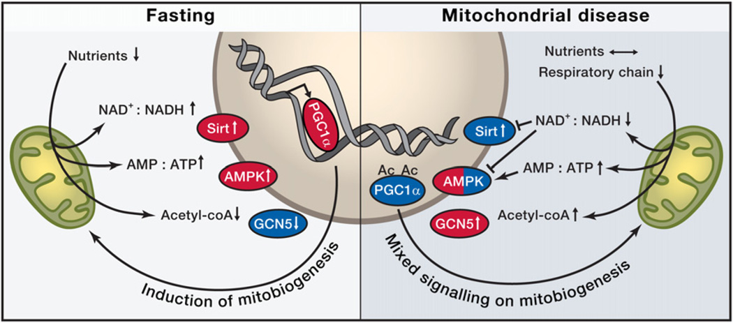
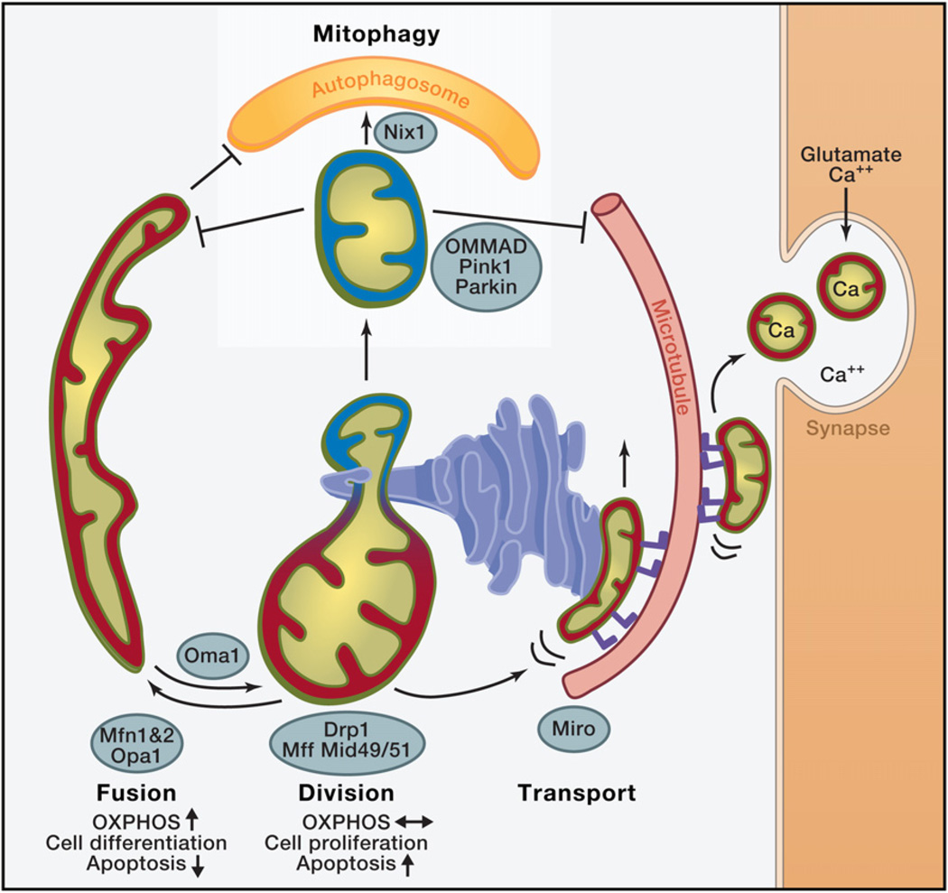
Mitochondria perform diverse yet interconnected functions, producing ATP and many biosynthetic intermediates while also contributing to cellular stress responses such as autophagy and apoptosis. Mitochondria form a dynamic, interconnected network that is intimately integrated with other cellular compartments. In addition, mitochondrial functions extend beyond the boundaries of the cell and influence an organism's physiology by regulating communication between cells and tissues. It is therefore not surprising that mitochondrial dysfunction has emerged as a key factor in a myriad of diseases, including neurodegenerative and metabolic disorders. We provide a current view of how mitochondrial functions impinge on health and disease.


Figures

Figure 1
Figure 1. Nutrient Sensors in Fasting and Their Roles in Mitochondrial Disease
Both fasting and mitochondrial disease can modify NAD+:NADH and AMP:ATP ratios through decreased nutrient availability or through reduced respiratory chain activity and have the potential to activate (red) nutrient sensors Sirtuin 1 (Sirt, an NAD+-dependent histone deacetylase) or AMP-activated kinase (AMPK) and increase mitochondrial biogenesis by activating peroxisome proliferator-activated receptor gamma coactivator 1-alpha (PGC-1alpha). Upon decreased utilization of acetyl-coenzyme A (acetyl-coA), GCN5 (lysine acetyltransferase 2A) is activated and acetylates PGC1alpha, to inactivate it (blue). NAD+, nicotinamide adenine dinucleotide, oxidized form; NADH, nicotinamide adenine dinucleotide, reduced form; AMP, adenosine monophosphate; ATP, adenosine triphosphate; Ac, acetyl group.
Figure 2
Figure 2. Roles of Mitochondrial Dynamics
Red: mitochondria with high membrane potential, with high oxidative phosphorylation (OXPHOS) activity. Blue: Mitochondria with low membrane potential. Mitofusin 1 or 2 (MFN1, MFN2) mediate mitochondrial outer-membrane fusion in a tissue-specific manner, and OPA1 (optic atrophy gene 1) mediates inner-membrane fusion. The zinc metalloprotease OMA1 proteolytically cleaves OPA1 under low membrane potential conditions, promoting fission. Mitochondrial dynamics factors 49 and 51 or mitochondrial fission factor (Mff) recruit DRP1 onto mitochondria at sites marked by endoplasmic reticulum tubules (ER), and DRP1 mediates mitochondrial division. In cultured cells, upon a decrease in mitochondrial membrane potential, PINK1 kinase recruits Parkin, a ubiquitin E3 ligase, which ubiquitinates several mitochondrial targets, including MFN1 and Miro, to facilitate the degradation of mitochondria via mitophagy. Parkin-mediated ubiquitination triggers OMMAD, outer-mitochondrial membrane-associated degradation—a proteosomal pathway that degrades ubiquinated OM proteins in a CDC48-dependent manner. OMMAD is probably cell type-dependent and may also function in quality control. In erythrocytes, mitophagy receptor Nix1 is involved in autophagosome recruitment. ER forms close contacts with mitochondria, essential for calcium regulation in cellular microcompartments. Miro (blue feet) is a mitochondrial receptor for kinesin via Milton that facilitates the transport of mitochondria on microtubules in a Ca2+-regulated manner. Upon synaptic activity in neurons, influx of glutamate and Ca2+ halts mitochondrial transport via Miro to position them at sites of synaptic activity that require Ca2+ uptake and ATP.
Figure 3
Figure 3. Interorganellar Communication
Ub, ubiquitin; red and blue, proteins in mitochondrial outer membrane are PINK1, a mitochondrial kinase, and the E3 ubiquitin ligase Parkin, recruited onto mitochondria by PINK1; MDV, mitochondria-derived vesicle; NAD, nicotinamide adenine dinucleotide, oxidized form; NADH, nicotinamide adenine dinucleotide, reduced form; AMP, adenosine monophosphate; ATP, adenosine triphosphate; ER, endoplasmic reticulum, tubules of which are marking sites of mitochondrial division; MAVS, mitochondrial antiviral signaling, which is activated by viral RNA; MAM, mitochondrial-associated endoplasmic reticulum membrane.
Figure 4
Figure 4. Organismal Effects of Mitochondrial Respiratory Chain Deficiency
Skeletal muscle interprets mitochondrial OXPHOS defect as a starvation response in the presence of normal nutrition. The defective muscle fibers secrete FGF21, a hormone-like cytokine, to blood circulation, mobilizing lipids from storage fat, affecting whole-organismal lipid metabolism as a chronic response.

References

    1. Ahlqvist KJ, Hämäläinen RH, Yatsuga S, Uutela M, Terzioglu M, Götz A, Forsström S, Salven P, Angers-Loustau A, Kopra OH, et al. Somatic progenitor cell vulnerability to mitochondrial DNA mutagenesis underlies progeroid phenotypes in Polg mutator mice. Cell Metab. 2012;15:100–109. - PubMed
    1. Ahola-Erkkilä S, Carroll CJ, Peltola-Mjösund K, Tulkki V, Mattila I, Seppänen-Laakso T, Oresic M, Tyynismaa H, Suomalainen A. Ketogenic diet slows down mitochondrial myopathy progression in mice. Hum. Mol. Genet. 2010;19:1974–1984. - PubMed
    1. Al Rawi S, Louvet-Vallée S, Djeddi A, Sachse M, Culetto E, Hajjar C, Boyd L, Legouis R, Galy V. Postfertilization autophagy of sperm organelles prevents paternal mitochondrial DNA transmission. Science. 2011;334:1144–1147. - PubMed
    1. Alexander C, Votruba M, Pesch UAE, Thiselton DL, Mayer S, Moore A, Rodriguez M, Kellner U, Leo-Kottler B, Auburger G, et al. OPA1, encoding a dynamin-related GTPase, is mutated in autosomal dominant optic atrophy linked to chromosome 3q28. Nat. Genet. 2000;26:211–215. - PubMed
    1. Anton F, Fres JM, Schauss A, Pinson B, Praefcke GJ, Langer T, Escobar-Henriques M. Ugo1 and Mdm30 act sequentially during Fzo1-mediated mitochondrial outer membrane fusion. J. Cell Sci. 2011;124:1126–1135. - PubMed

Publication types