. 2012 Mar 16:9:55.
doi: 10.1186/1742-2094-9-55.

Leukotrienes inhibit early stages of HIV-1 infection in monocyte-derived microglia-like cells

Affiliations

Leukotrienes inhibit early stages of HIV-1 infection in monocyte-derived microglia-like cells

Jonathan Bertin et al. J Neuroinflammation. .

Abstract

Background: Microglia are one of the main cell types to be productively infected by HIV-1 in the central nervous system (CNS). Leukotriene B4 (LTB4) and cysteinyl-leukotrienes such as LTC4 are some of the proinflammatory molecules produced in infected individuals that contribute to neuroinflammation. We therefore sought to investigate the role of leukotrienes (LTs) in HIV-1 infection of microglial cells.

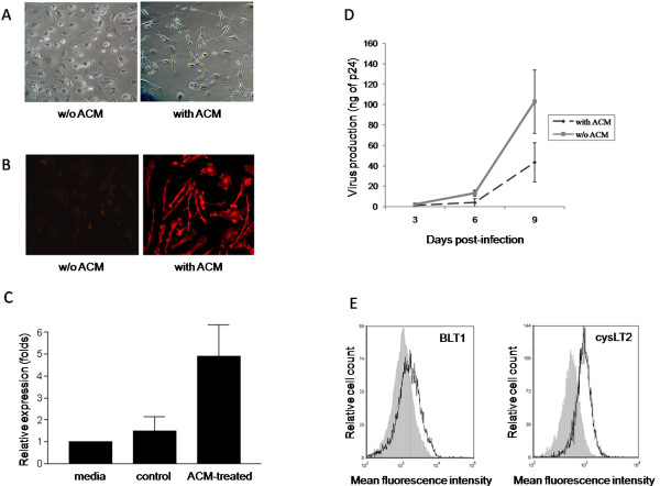
Methods: To evaluate the role of LTs on HIV-1 infection in the CNS, monocyte-derived microglial-like cells (MDMis) were utilized in this study. Leukotriene-treated MDMis were infected with either fully replicative brain-derived HIV-1 isolates (YU2) or R5-tropic luciferase-encoding particles in order to assess viral production and expression. The efficacy of various steps of the replication cycle was evaluated by means of p24 quantification by ELISA, luciferase activity determination and quantitative real-time polymerase chain reaction (RT-PCR).

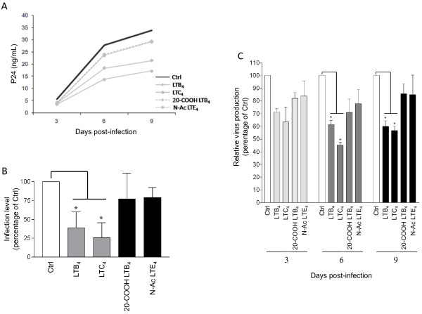
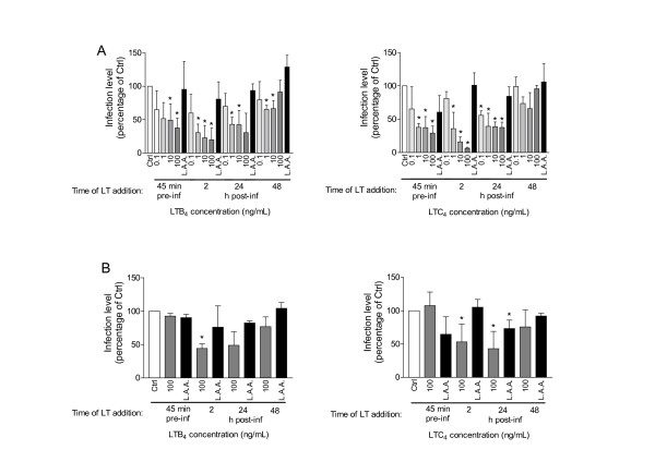
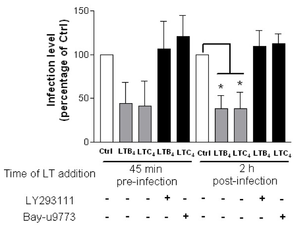
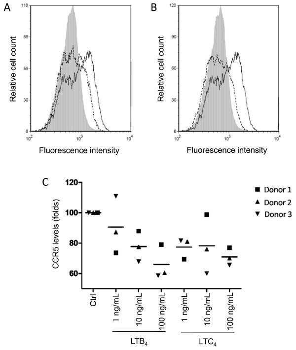
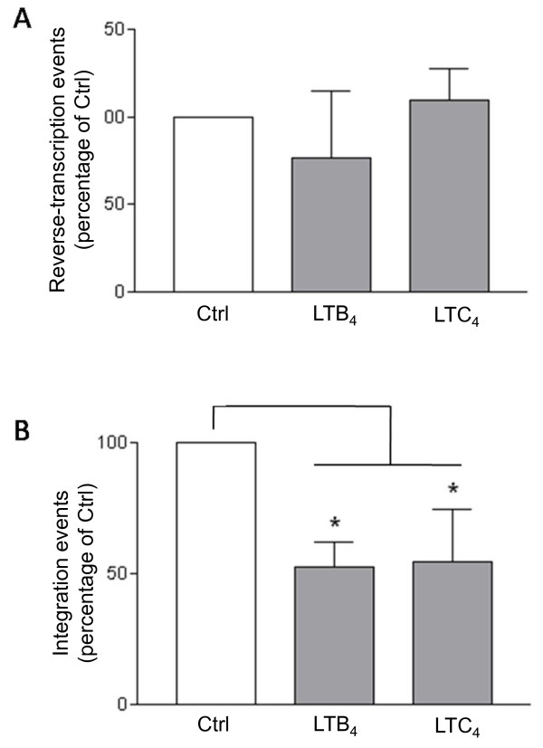
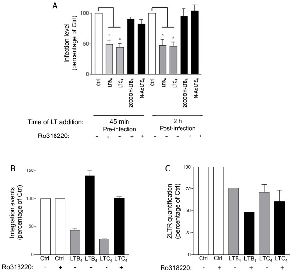
Results: We report in this study that virus replication is reduced upon treatment of MDMis with LTB4 and LTC4. Additional experiments indicate that these proinflammatory molecules alter the pH-independent entry and early post-fusion events of the viral life cycle. Indeed, LT treatment induced a diminution in integrated proviral DNA while reverse-transcribed viral products remained unaffected. Furthermore, decreased C-C chemokine receptor type 5 (CCR5) surface expression was observed in LT-treated MDMis. Finally, the effect of LTs on HIV-1 infection in MDMis appears to be mediated partly via a signal transduction pathway involving protein kinase C.

Conclusions: These data show for the first time that LTs influence microglial cell infection by HIV-1, and may be a factor in the control of viral load in the CNS.


Figures

Figure 1
Figure 1
Characterization of microglial-like cells. Monocytes were isolated from peripheral blood mononuclear cells (PBMCs) of healthy donors, and then cultured either in absence or presence of astrocyte-conditioned medium (ACM). (A) Cell morphology was observed by phase-contrast microscopy. Monocytes cultured in the absence of ACM were used as control cells. The results shown are representative of at least three different donors. (B) ACM-differentiated cells were permeabilized and stained with anti-ionized calcium binding adaptor protein-1 (IBA1) antibodies. IBA1 expression was observed by epifluorescence microscopy. The results shown are representative of at least three distinct donors. (C) Substance P secretion by monocyte-derived microglial-like cells (MDMis) and non-ACM-treated control cells was evaluated by slot-blot. Results from two independent donors (means ± SEM) are presented. (D) ACM-treated and non-treated monocytes were infected with YU2. Virus replication was assessed by monitoring the p24 content at days 3, 6 and 9 post infection. The means ± SEM are calculated from three independent experiments with triplicate samples. (E) The surface expression of BLT1 and cysLT2 receptors on ACM-treated monocytes was determined by flow cytometry. The results shown represent a single donor out of a total of four.
Figure 2
Figure 2
Leukotrienes (LTs) reduce susceptibility of monocyte-derived microglial-like cells (MDMis) to HIV-1 infection. (A) MDMis were pretreated for 45 minutes with 100 ng/ml of the indicated LTs prior to infection with YU2. At 2 h after infection, cells were then thoroughly washed and incubated in fresh media containing the listed LTs. Additional LTs were also replenished at days 3 and 6 post infection. Viral production was monitored at 3, 6 and 9 days post infection by measuring the p24 content. The results shown are from one representative donor of a total of three. (B) The p24 values for all three donors tested are depicted as percentage of untreated control to compensate for donor-to-donor virus infection variability. (C) Production of infectious virus particles by LT-treated MDMis was quantified at 9 days post infection in cell-free supernatants using the TZM-bl indicator cell line. Results shown represent mean luciferase activity ± SD calculated from three independent experiments with triplicate samples and are expressed as the relative percentage of infectious virus production with respect to non-treated cells (*P < 0.05).
Figure 3
Figure 3
Leukotrienes (LTs) exert an inhibitory effect on the early stages of HIV-1 infection in monocyte-derived microglial-like cells (MDMis). Cells were either left untreated, or treated with increasing concentrations of LTB4 (left panels) or LTC4 (right panels), either 45 minutes before virus exposure or 2 h to 48 h post infection, as indicated. Cells were first washed extensively with phosphate-buffered saline (PBS) and then infected at 45 minutes after addition of LTs. MDMis were washed a second time at 2 h after infection before adding LTs. No washes were carried out before adding LTs at 24 h and 48 h. Infection was carried out with a luciferase encoding reporter virus pseudotyped with JR-FL (A) or vesicular stomatitis virus envelope glycoprotein G (VSV-G) envelope (B). The less active analogs (called LAA) 20-carboxy-LTB4 and N-acetyl LTE4 were used as negative controls for LTB4 and LTC4, respectively. The extent of virus infection was estimated by measuring virus-encoded luciferase activity at 7 days post infection. The results shown represent the means ± SD calculated from three, or four (bottom right panel), independent experiments with triplicate samples and are expressed as the percentage of luciferase activity with respect to non-treated cells (*P < 0.05).
Figure 4
Figure 4
Leukotriene (LT) receptor antagonists block the modulatory effect of LTB4 and LTC4 on HIV-1 replication in monocyte-derived microglial-like cells (MDMis). Cells were either left untreated (Ctrl) or treated with the listed LT receptor antagonists (that is, LY293111 or Bay-u9773) for 45 minutes before being thoroughly washed. MDMis were then either exposed to LTs 45 minutes before or 2 h following HIV-1 infection, after which cells were thoroughly washed. Virus infection was carried out using JR-FL pseudotyped viruses. The extent of virus infection was estimated by measuring virus-encoded luciferase activity at 7 days post infection. The results shown represent the means ± SD calculated from three independent experiments with triplicate samples and are expressed as the percentage of luciferase activity with respect to non-treated cells (*P < 0.05).
Figure 5
Figure 5
Leukotriene (LT)B4 and LTC4 induce a downmodulation of C-C chemokine receptor type 5 (CCR5) surface expression on monocyte-derived microglial-like cells (MDMis). Cells were either left untreated (continuous lines) or treated with 10 ng/ml of (A) LTB4 or (B) LTC4 (dotted lines) for 24 h before labeling with a phycoerythrin (PE)-conjugated anti-CCR5 antibody or PE-conjugated irrelevant control antibody (gray areas). Expression levels of CCR5 were determined by flow cytometry. Results shown are from one donor representative of three. (C) MDMis were either left untreated or treated with increasing concentrations of LTs for 24 h before labeling with a PE-conjugated anti-CCR5 antibody. To compensate for donor-to-donor variations, the results shown represent the mean percentage of CCR5 positive cells ± SD calculated from three independent experiments and are expressed as percentages of control (Ctrl) (*P < 0.05).
Figure 6
Figure 6
Leukotrienes (LTs) have no impact on HIV-1 reverse transcripts, whereas they affect viral integration in monocyte-derived microglial-like cells (MDMis). Cells were first exposed to YU2 for 2 h. MDMis were then either left untreated (Ctrl) or treated with 10 ng/ml of LTB4 or LTC4. DNA was then extracted 8 h post infection for monitoring reverse transcribed products or 24 h post infection for assessing integration events. Quantification was achieved by real-time polymerase chain reaction (PCR). Amplified products were normalized using the housekeeping β-globin gene. To compensate for donor-to-donor variations, the results shown represent the means ± SD calculated from three independent experiments and are expressed as percentages of control (Ctrl) (*P < 0.05).
Figure 7
Figure 7
Leukotrienes (LTs) reduce HIV-1 infection of monocyte-derived microglial-like cells (MDMis) in a protein kinase C (PKC)-dependent manner. (A) MDMis were first either left untreated or treated with the large-spectrum PKC inhibitor Ro318220 (1 μM) for 30 minutes. LTs (10 ng/ml) were then added to cells either for 45 minutes before infection or after 2 h of incubation with JR-FL pseudotypes. Luciferase activity was measured 7 days post infection. (B, C) MDMis were first infected with vesicular stomatitis virus envelope glycoprotein G (VSV-G) pseudotypes for a 2 h period, cells were then treated with Ro318220 for 45 minutes, washed, and treated with LTs (100 ng/ml). Total DNA was extracted from cells in order to quantify HIV-1 integration events (panel B) and nuclear 2LTR circles (C) by real-time polymerase chain reaction (PCR). The results shown are expressed as percentages of control (that is, untreated cells) and calculated from five (A) or two (B, C) independent experiments in triplicate samples (*P < 0.05).

References

    1. Antinori A, Arendt G, Becker JT, Brew BJ, Byrd DA, Cherner M, Clifford DB, Cinque P, Epstein LG, Goodkin K, Gisslen M, Grant I, Heaton RK, Joseph J, Marder K, Marra CM, McArthur JC, Nunn M, Price RW, Pulliam L, Robertson KR, Sacktor N, Valcour V, Wojna VE. Updated research nosology for HIV-associated neurocognitive disorders. Neurology. 2007;69:1789–1799. doi: 10.1212/01.WNL.0000287431.88658.8b. - DOI - PMC - PubMed
    1. Ghafouri M, Amini S, Khalili K, Sawaya BE. HIV-1 associated dementia: symptoms and causes. Retrovirology. 2006;3:28. doi: 10.1186/1742-4690-3-28. - DOI - PMC - PubMed
    1. Canestri A, Lescure FX, Jaureguiberry S, Moulignier A, Amiel C, Marcelin AG, Peytavin G, Tubiana R, Pialoux G, Katlama C. Discordance between cerebral spinal fluid and plasma HIV replication in patients with neurological symptoms who are receiving suppressive antiretroviral therapy. Clin Infect Dis. 2010;50:773–778. doi: 10.1086/650538. - DOI - PubMed
    1. Christo PP, Greco DB, Aleixo AW, Livramento JA. HIV-1 RNA levels in cerebrospinal fluid and plasma and their correlation with opportunistic neurological diseases in a Brazilian AIDS reference hospital. Arq Neuropsiquiatr. 2005;63:907–913. - PubMed
    1. De Luca A, Ciancio BC, Larussa D, Murri R, Cingolani A, Rizzo MG, Giancola ML, Ammassari A, Ortona L. Correlates of independent HIV-1 replication in the CNS and of its control by antiretrovirals. Neurology. 2002;59:342–347. - PubMed

Publication types