. 2012 Apr 10;22(7):575-82.
doi: 10.1016/j.cub.2012.02.044. Epub 2012 Mar 15.

A role for the centrosome and PAR-3 in the hand-off of MTOC function during epithelial polarization

Affiliations

A role for the centrosome and PAR-3 in the hand-off of MTOC function during epithelial polarization

Jessica L Feldman et al. Curr Biol. .

Abstract

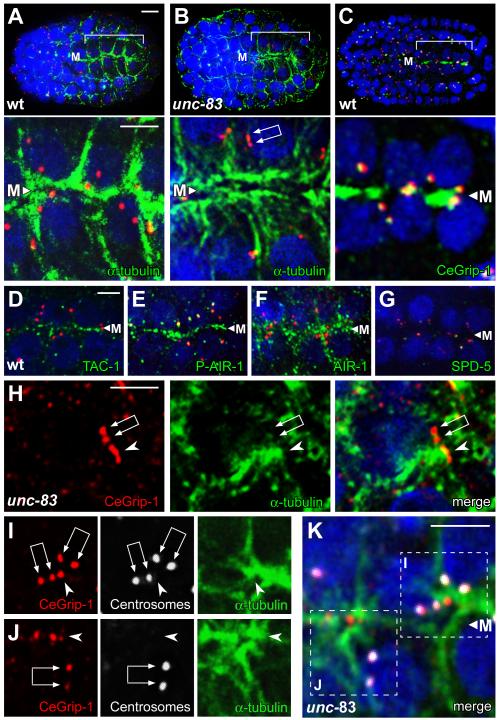
Background: The centrosome is the major microtubule organizing center (MTOC) in dividing cells and in many postmitotic, differentiated cells. In other cell types, however, MTOC function is reassigned from the centrosome to noncentrosomal sites. Here, we analyze how MTOC function is reassigned to the apical membrane of C. elegans intestinal cells.

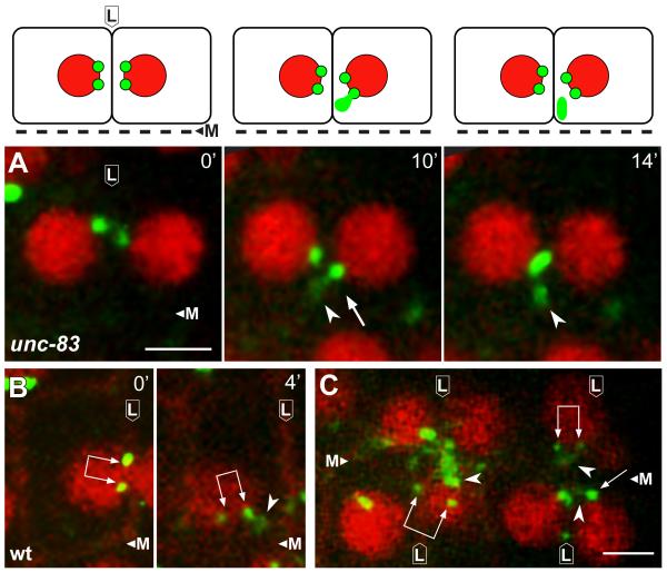
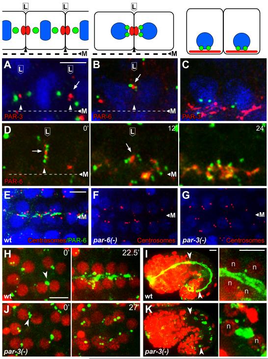
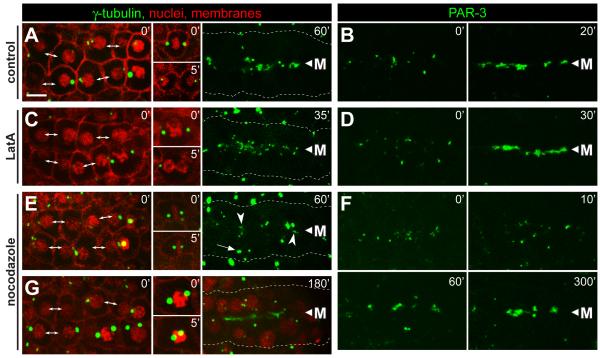
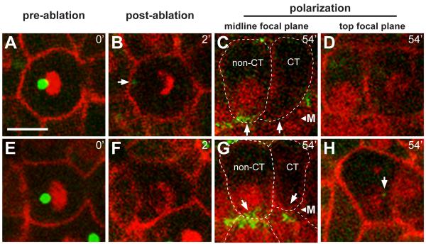
Results: After the terminal intestinal cell division, the centrosomes and nuclei move near the future apical membranes, and the postmitotic centrosomes lose all, or nearly all, of their associated microtubules. We show that microtubule-nucleating proteins such as γ-tubulin and CeGrip-1 that are centrosome components in dividing cells become localized to the apical membrane, which becomes highly enriched in microtubules. Our results suggest that centrosomes are critical to specify the apical membrane as the new MTOC. First, γ-tubulin appears to redistribute directly from the migrating centrosome onto the lateral then apical membrane. Second, γ-tubulin fails to accumulate apically in wild-type cells following laser ablation of the centrosome. We show that centrosomes localize apically by first moving toward lateral foci of the conserved polarity proteins PAR-3 and PAR-6 and then move together with these foci toward the future apical surface. Embryos lacking PAR-3 fail to localize their centrosomes apically and have aberrant localization of γ-tubulin and CeGrip-1.

Conclusions: These data suggest that PAR proteins contribute to apical polarity in part by determining centrosome position and that the reassignment of MTOC function from centrosomes to the apical membrane is associated with a physical hand-off of nucleators of microtubule assembly.


Figures

Figure 1
Figure 1
Centrosomes and γ-tubulin localization during MTOC reassignment (A-C) Images from live recording of the E16 primordium, showing sister E16 cells from birth through polarization. Reporters show γ-tubulin (green, γ-tubulin:GFP), nuclei (red, histone:mCherry) and membranes (red, membrane:Cherry). Cells are oriented with respect to the midline (M) as diagrammed in the cartoons beneath each panel. Note the transition from round to columnar cell morphology associated with polarization. (D-F) Electron micrographs of centrosomes from stages corresponding to panels A, B, and C, respectively. Note the decrease in diameter of the PCM (indicated with purple arrows in panels D and E) as the nucleus (“nuc”) and centrosome localizes near the lateral membrane (L), and the apparent absence of PCM as they move near the midline in panel F. Green arrowheads indicate examples of microtubules contacting the PCM. Scale bar (A-C, 5μm, D-F, 200nm). See also Figure S1 and Movie S1.
Figure 2
Figure 2
Generation of the midline/apical MTOC (A-K) All panels are optical sections through the midline (M) of an E16 primordium, immunostained as indicated; nuclei are stained with DAPI (blue). Centrosome pairs appear as two closely paired dots with variable spacing (double arrows in panels B, H, I, and J), or are unresolved as a larger dot. The upper panels in A-C show low magnification views of the entire embryo with the primordium bracketed, and the lower panels show high magnification views of same E16 cells; centrosomes are stained with IFA1 (red). In the unc-83 mutant (B), note the microtubules extending between the apical membrane and the nucleus. (C-F) The PCM proteins CeGrip-1, TAC-1, and AIR-1 are enriched at the midline/future apical surface. Although the kinase AIR-1 primarily co-localizes with microtubules during this stage, the phosphorylated, active form of AIR-1 is restricted to the apical surface and centrosomes (compare E and F; [30]). Panels H-J show examples of plumes (arrowheads) of CeGrip-1 near the centrosome pair; panels I and J are color separated images of the merged, boxed regions in K. Note the concentration of α-tubulin by the plume of CeGrip-1, and the relative absence of α-tubulin by the centrosome pair. Scale bar = 5μm (A-C), 2.5μm (D-K and insets in A-C). See also Figure S2, Figure S3, and Movie S2.
Figure 3
Figure 3
A plume of γ-tubulin appears to emanate from the centrosome during MTOC reassignment Panels are from live recordings of the E16 primordium in wild-type or unc-83 mutant embryos; lateral membranes (L) and midline (M) are indicated. Reporters show γ-tubulin (green, γ-tubulin:GFP), nuclei (red, histone:mCherry), and membranes (B and C, red, membrane:Cherry). Centrosome pairs are indicated by single or double arrows, depending on whether one or both centrosomes is visible in the frame. Note plumes of γ-tubulin (arrowheads) associated with centrosomes. Images in A are diagrammed above each panel. Scale bar (2.5 μm). See also Movie S3.
Figure 4
Figure 4
PAR proteins in centrosome positioning and γ-tubulin localization (A-C) Intestinal primordial cells in immunostained embryos at (A) metaphase of the E8 to E16 division, (B) shortly after division when paired centrosomes migrate to lateral membranes, and (C) during apical polarization; cartoons of the cells are shown in the upper panels. The images show centrosomes (green; SPD-5 in panel A and IFA1 in, B, C), and PAR-3 or PAR-6 as indicated; nuclei are stained with DAPI (blue). Note that the paired centrosomes in panel B have moved toward the lateral focus of PAR-6. (D) Image sequence from live E16 cells beginning at a stage similar to panel B and showing centrosomes (green, γ-tubulin:GFP) and PAR-6 (red, PAR-6:Cherry); time in minutes at upper right. Note that the centrosome pairs in both cells move apically with the focus of PAR-6, and that PAR-6 spreads after reaching the apical surface. (E-G) Images of the E16 primordium in immunostained wild-type embryos and embryos depleted of both maternal and zygotic PAR-6 or PAR-3 (n=25 and 27 embryos, respectively). Note that most of the paired centrosomes (red, IFA) are apical after depletion of PAR-6 but not after depletion of PAR-3. (H-K) γ-tubulin (green, γ-tubulin:GFP) and nuclei (red, histone:Cherry, ‘n’) in wild type embryos (H,I) and embryos depleted of PAR-3 (J,K). Note the failure of centrosomes and γ-tubulin to localize apically in the PAR-3-depleted embryo. The left panels in I and K show low magnifications of entire embryos during later morphogenesis, with the intestines indicated by arrowheads; the right panels show high magnification views of some of the intestinal cells. γ-tubulin is apical in the wild-type embryo, but ectopic in the PAR-3-depleted embryo. Scale bar (A-C, 2.5μm, E-K, 5μm). See also Figure S4, Movie S4, and Movie S5.
Figure 5
Figure 5
Inhibition of microfilaments and microtubules during epithelial polarization. Each column shows examples of live, E16 primordia from wild-type embryos expressing the reporters indicated at top; the reporters used are as in Figure 1. Embryos were either mock treated (control), or exposed to latrunculin (LatA, 10μM) or nocodazole (10μg/mL) as indicated. The left panels for each column show the primordium immediately after exposure to the inhibitor. At the stage selected for γ-tubulin analysis, the primordium includes newly separated sister cells (double-headed arrows), as well as some cells that have not finished the E8 to E16 division; these latter cells arrest division (A,E,G) or fail cytokinesis (C) after treatment with inhibitor (inset panels). In the stage selected for PAR-3 analysis, PAR-3 foci were visible on the lateral membranes of E16 cells (left panels, t=0). (A-D) In control embryos and LatA-treated embryos, centrosomes, γ-tubulin and PAR-3 show robust apical localization before 60 minutes (see also timed sequence in Figure 4H). In LatA-treated cells that failed cytokinesis, γ-tubulin did not localize to the apical surface within the same time period (data not shown). (E-G) Embryos treated with nocodazole show only irregular apical localization by 60 minutes, but much better localization of γ-tubulin and PAR-3 by 180 minutes and 200 minutes, respectively. Centrosomes (E, 60′ arrow, G) and nuclei (G) fail to move apically following nocodazole treatment. Note the spreading of PAR-3 foci away from the midline 10 minutes after nocodazole treatment, suggesting that these foci normally traffic toward the midline on microtubules. Scale bar = 5μm. See also Movie S6.
Figure 6
Figure 6
Centrosomes are required for accumulation of γ-tubulin at the apical surface Live images of two wild type embryos (A-D and E-H) before (t=0) and after (t=2 minutes) laser ablation of the centrosome, and again after 54 minutes; reporters as in Figure 1. γ-tubulin normally accumulates at the midline before 1 hour, as is visible in the non-centrosome ablated (non-CT) cells, but has not accumulated in the cells with the ablated centrosomes (CT). Note that the nuclei in CT cells have not localized to the midline, but are instead at different focal planes (top focal plane, D and H). The arrow in panel H indicates a non-apical, small cytoplasmic focus of γ-tubulin that likely corresponds to a fragment of the ablated centrosome. CT cells have levels of cytoplasmic γ-tubulin that are comparable to non-CT cells, but lack apical accumulation; the apical surface of CT cells showed a statistically insignificant enrichment of γ-tubulin (−.0018±.012, n=19) compared to non-CT cells (.2081±.082, n=38, two tailed t-test; p ≤ 1.83×10⁻¹⁷). Scale bar = 5μm. See also Movie S7.

Comment in

References

    1. Hannak E, et al. The kinetically dominant assembly pathway for centrosomal asters in Caenorhabditis elegans is gamma-tubulin dependent. J Cell Biol. 2002;157:591–602. - PMC - PubMed
    1. Bray D, Bunge MB. Serial analysis of microtubules in cultured rat sensory axons. J Neurocytol. 1981;10:589–605. - PubMed
    1. Baas PW, Deitch JS, Black MM, Banker GA. Polarity orientation of microtubules in hippocampal neurons: uniformity in the axon and nonuniformity in the dendrite. Proc Natl Acad Sci U S A. 1988;85:8335–9. - PMC - PubMed
    1. Tassin AM, Maro B, Bornens M. Fate of microtubule-organizing centers during myogenesis in vitro. J Cell Biol. 1985;100:35–46. - PMC - PubMed
    1. Bré MH, Kreis TE, Karsenti E. Control of microtubule nucleation and stability in Madin-Darby canine kidney cells: the occurrence of noncentrosomal, stable detyrosinated microtubules. J Cell Biol. 1987;105:1283–96. - PMC - PubMed

Publication types

MeSH terms