Review
. 2012 Apr;18(4):224-32.
doi: 10.1016/j.molmed.2012.02.001. Epub 2012 Mar 16.

Two opposing roles of O-glycans in tumor metastasis

Affiliations
Review

Two opposing roles of O-glycans in tumor metastasis

Shigeru Tsuboi et al. Trends Mol Med. 2012 Apr.

Abstract

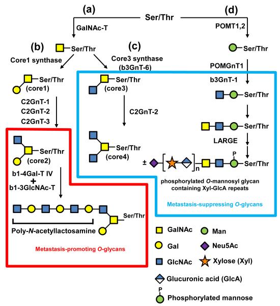
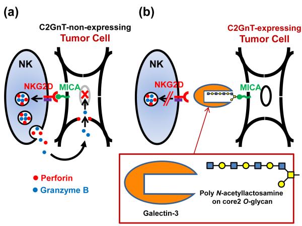
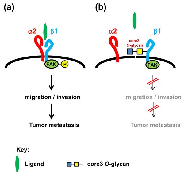
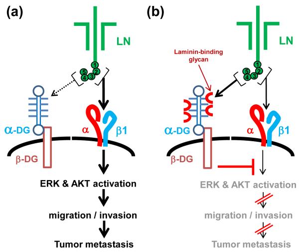
Despite the high prevalence of metastatic cancers and the poor outcome for patients, the processes of tumor metastasis still remain poorly understood. It has been shown that cell-surface carbohydrates attached to proteins through the amino acids serine or threonine (O-glycans) are involved in tumor metastasis, with the roles of O-glycans varying depending on their structure. Core2 O-glycans allow tumor cells to evade natural killer (NK) cells of the immune system and survive longer in the circulatory system, thereby promoting tumor metastasis. Core3 O-glycans or O-mannosyl glycans suppress tumor formation and metastasis by modulating integrin-mediated signaling. Here, we highlight recent advances in our understanding of the detailed molecular mechanisms by which O-glycans promote or suppress tumor metastasis.


Figures

Figure 1
Figure 1. O-glycan biosynthesis pathways that promote or suppress tumor metastasis
(a) To begin synthesis of core2 and core4 O-glycans N-acetylgalactosamine (GalNAc) is transferred to a serine (Ser) or threonine (Thr) residue in a polypeptide by the peptide GalNAc transferase (GalNAc-T), as shown on the left side of the figure. GalNAcα1-Ser/Thr is then converted by core1 synthase to Galβ1-3GalNAcα1-Ser/Thr (core1) (b). Core1 is converted to core2 by core2 β-1,6-N-acetylglucosaminyltransferase (C2GnT-1, −2 and −3). β-1,4-galactosidase IV (β1-4Gal-T IV) together with β-1,3-N-acetylglucosaminyltransferase (β1-3GlcNAc-T V) synthesizes poly-N-acetyllactosamine on core2 O-glycans. The Number of Galβ1-4GlcNAc disaccharide unit repeat varies depending on carrier molecules and cell types. (c) GalNAcα1-Ser/Thr is also converted by core3 synthase (β3GnT-6) to core3. Core3 is then converted by C2GnT-2 to core4. By contrast, synthesis of O-mannosyl glycans begins with transfer of mannose to a serine or threonine residue by protein O-mannosyltransferase 1 and 2 (POMT1 and 2) (d). LARGE has α-xylosyltransferase and β-glucuronic acid transferase activities. Manα1-Ser/Thr is converted by a series of glycosyltransferases, including protein O-mannoseβ-1,2-N-acetylglucosaminyltransferase 1 (POMGnT1), β3GnT-1, and LARGE, to O-mannosyl glycan, which contains sialic acid (Neu5Ac), α-xylose. and β-glucuronic acid disaccharide unit repeats and phosphorylated mannose. As discussed in the main text, poly-N-acetyllactosamine-modified core2 O-glycans can be described as “Metastasis-promoting O-glycans” (bounded by the red line) and core3 and core4-containing O-glycans and phosphorylated O-mannosyl glycans can be described as “Metastasis-suppressing O-glycans” (bounded by the blue line).
Figure 2
Figure 2. Core2 O-glycans promote tumor metastasis by evading NK cell attack
The molecular mechanism by which C2GnT-expressing tumor cells evade NK cells. (a) On the left is an illustration of a major mechanism by which tumors are rejected by NK cells. NK cells are activated by the interaction of NKG2D with MICA on the surface of the tumor cells. Activated NK cells secrete perforin and granzyme B to induce apoptosis of target tumor cells. (b) On the right is the mechanism by which C2GnT-expressing tumor cells evade NK cell attack. The protein galectin-3 binds to poly-N-acetyllactosamine in the NKG2D-binding site of MICA. Galectin-3 reduces the affinity of MICA for NKG2D, severely impairing NK cell activation. This masking of the tumor cells increases survival of the tumor cells in host blood circulation, promoting tumor metastasis.
Figure 3
Figure 3. The role of core3 O-glycan in suppressing migration and tumor metastasis
(a) In tumor cells with low core3 synthase expression, ligand binding to α2β1 integrin induces phosphorylation of FAK. Phosphorylated FAK transduces signals to mediate tumor cell migration and invasion, promoting metastasis. (b) In cells that express core3 synthase, core3 O-glycans on the β1 subunit of α2β1 integrin prevent the association of the α2 and β1 subunits, reducing the amount of functional α2β1 complex on the cell-surface. The reduced levels of α2β1 limit FAK phosphorylation, impairing signal transduction. Impaired signaling reduces cell migration and invasion and suppresses tumor formation and metastasis.
Figure 4
Figure 4. The role of phosphorylated O-mannosyl glycan (laminin-binding glycan) in suppressing tumor metastasis
(a) In tumor cells with downregulated β3GnT1 expression, laminin (LN) globular domains 4 and 5 are unable to bind to α-DG because the α-DG laminin-binding glycan is not synthesized (denoted by broken arrow), but laminin globular domains 1, 2, and 3 still bind to β1 integrin (denoted by bold and solid arrow), activating ERK-AKT signaling. This activated signaling mediates high tumor migration and invasion, resulting in metastasis. (b) In β3GnT1-expressing tumor cells, laminin-binding glycan containing α-xylose and β-glucuronic acid disaccharide unit repeats and phosphorylated mannose is synthesized on α-DG. Laminin globular domains 4 and 5 bind to α-DG through laminin-binding glycan (denoted by solid arrow), decreasing the amount of laminin available to bind β1 integrin (denoted by thin and solid arrows) and counteracting the ERK-AKT signaling. This results in suppressed tumor migration and metastasis. The globular domains 1 through 5 of laminin α-chain are indicated by the circled numbers.

Similar articles

Cited by

References

    1. Fuster MM, Esko JD. The sweet and sour of cancer: glycans as novel therapeutic targets. Nat Rev Cancer. 2005;5:526–542. - PubMed
    1. Dennis JW, et al. UDP-N-acetylglucosamine:alpha-6-D-mannoside beta1,6 N-acetylglucosaminyltransferase V (Mgat5) deficient mice. Biochim Biophys Acta. 2002;1573:414–422. - PubMed
    1. Yoshimura M, et al. Suppression of lung metastasis of B16 mouse melanoma by N-acetylglucosaminyltransferase III gene transfection. Proc Natl Acad Sci U S A. 1995;92:8754–8758. - PMC - PubMed
    1. Suzuki-Anekoji M, et al. HNK-1 Glycan Functions as a Tumor Suppressor for Astrocytic Tumor. J Biol Chem. 2011;286:32824–32833. - PMC - PubMed
    1. Brockhausen I. Mucin-type O-glycans in human colon and breast cancer: glycodynamics and functions. EMBO Rep. 2006;7:599–604. - PMC - PubMed

Publication types